Description
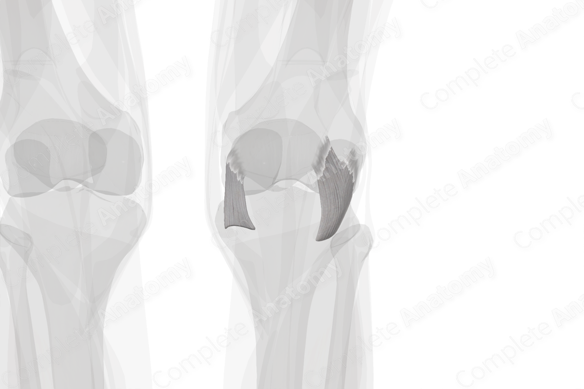
The patellofemoral joint is a part of the knee joint which is subject to a variety of forces by surrounding muscles, tendons, and ligaments. In this context, the patellar retinacula serve as important structures to provide stability to the patellofemoral joint throughout the wide range of knee motion.
While the tendon of the quadriceps muscle extends down on either side of the patella, to insert into the tibial tuberosity, the patellar retinacula (medial and lateral) extend obliquely and transversely as delicate, layered, fibrous connective tissue expansions to the patellar margin. These are primarily the extensions of vastus medialis and lateralis muscles.


Related parts of the anatomy
