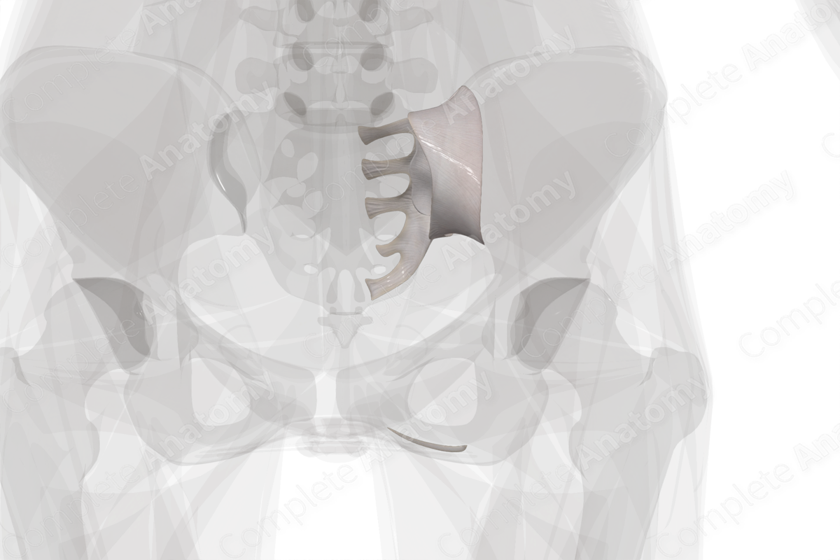
Sacroiliac Joint (Left) Structure
The right and left sacroiliac joints connect the articular surfaces of the ilium and the lateral sacrum. In children, this joint is synovial and permits limited movement. As age increases, this joint becomes a syndesmotic and may even ossify in older individuals.
Related parts of the anatomy
Sacroiliac Joint (Left) Anatomical Relations
Anterior to the sacroiliac joint, the external and internal iliac veins unite to form the common iliac veins, in addition to the bifurcation of the common iliac arteries at this level. The piriformis muscle attaches to the anterior aspect of the joint capsule and separates the joint from the sacral plexus.
Sacroiliac Joint (Left) Function
The sacroiliac joints connect the vertebral column to the pelvis and is one of the most stable joints in the body. It permits weight transmission from the vertebral column to the pelvis.
Learn more about this topic from other Elsevier products
Sacroiliac Joint

It is a term used to describe pain in the sacroiliac region related to abnormal sacroiliac joint movement or alignment.


