Description
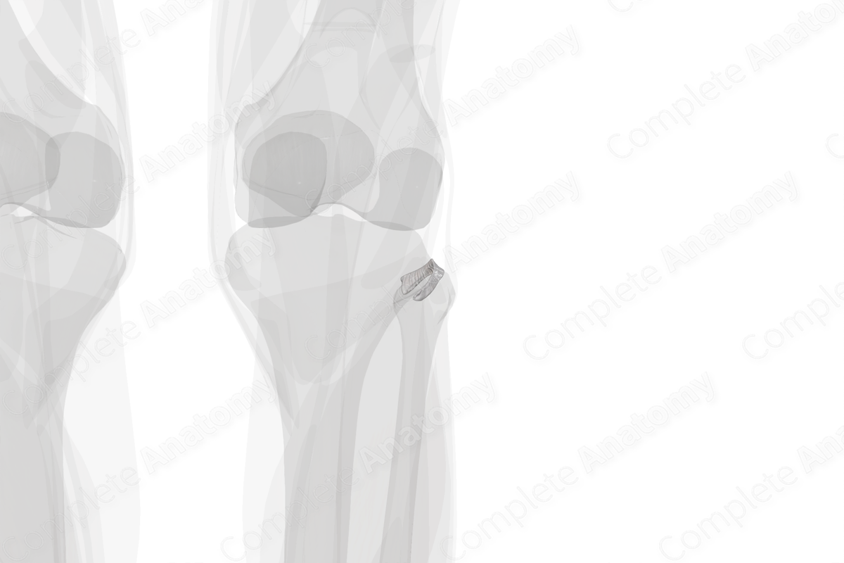
The superior tibiofibular joint is the articulation between the lateral condyle of the tibia and the head of the fibula. The superior tibiofibular joint is a plane, synovial, gliding joint and its surfaces are covered by hyaline cartilage. Unlike the inferior tibiofibular joint, it has a fibrous capsule that is attached to the borders of the articular surfaces (Standring, 2016). The fibrous capsule of the superior tibiofibular joint is thick anteriorly and posteriorly and is strengthened by the anterior and posterior ligaments of the fibular head and the interosseous membrane of the leg (Wineski, 2018). The function of the fibrous capsule, along with the ligaments, is to maintain the stability of the superior tibiofibular joint.
The superior tibiofibular joint receives its vascular supply from the anterior and posterior tibial recurrent arteries. It receives sensory innervation from the crural interosseous nerve and by recurrent articular branch of the common fibular nerve (Standring, 2016).


Related parts of the anatomy
References
Standring, S. (2016) Gray's Anatomy: The Anatomical Basis of Clinical Practice. Gray's Anatomy Series 41st edn.: Elsevier Limited.
Wineski, L. E. (2018) Snell's Clinical Anatomy by Regions. 10 edn.: Wolters Kluwer Health.
