Description
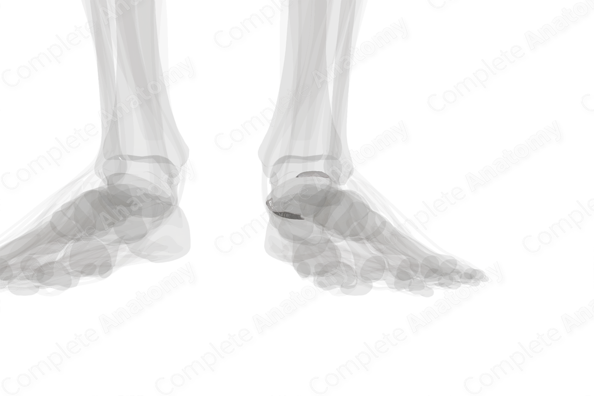
The talocalcaneonavicular joint is formed by the articulation of the talus, calcaneus, and navicular bones. The talus forms a continuous concave articular surface that receives the:
—calcaneus via the anterior and middle facets;
—navicular bone via the navicular articular facet.
The talocalcaneal part of the talonavicular joint can be classified as part of the anatomical subtalar joint, thus named the surgical subtalar joint. The talonavicular part of the talocalcaneonavicular joint is considered part of the transverse tarsal joint.
The talocalcaneonavicular joint is a modified multiaxial joint and plays a role in movements such as gliding and rotation. The bones are held together by a fibrous capsule, strengthened by the talonavicular and plantar calcaneonavicular ligaments. Additionally, the bifurcate ligament also contributes to the stability of this joint.


Related parts of the anatomy
