Description
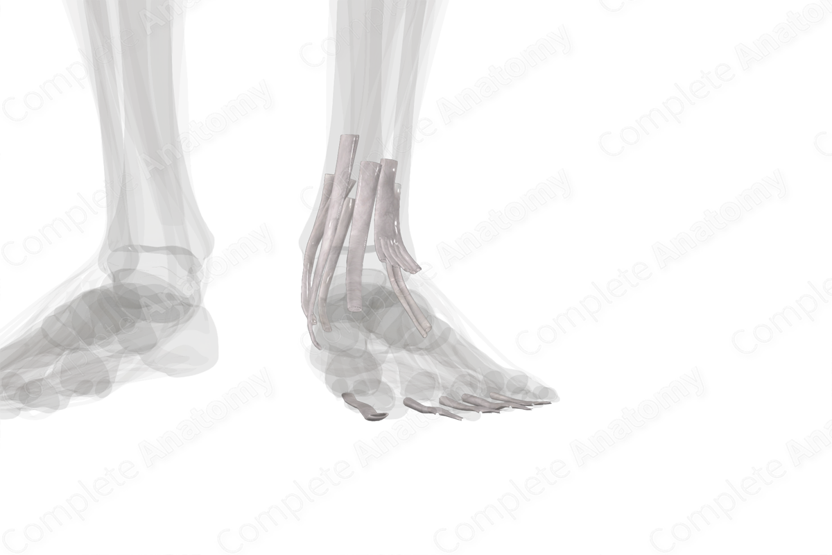
Several tendons originating from muscles in the leg traverse the ankle to enter the foot. They pass beneath retinacula which holds the tendons in place, preventing subluxation. These tendons are covered by modified bursa, called tendon sheaths, which minimize friction with the surrounding structures. The sheaths, composed of both fibrous and synovial layers, wrap around the tendons, ensuring that the tendons can glide over adjacent structures and allow for minimal friction during movement. Additionally, the tendon sheaths produce synovial fluid, ensuring that the tendons are well lubricated.
The tendon sheaths of the lower limb can be categorized according to their relation at the ankle joint.
The anterior tarsal tendinous sheaths include are found anterior to the ankle joint and include the:
—tendon sheath of tibialis anterior muscle;
—tendon sheath of extensor hallucis longus muscle;
—tendon sheath of extensor digitorum longus muscle.
The tibial tarsal tendon sheaths are located posteromedial to the ankle joint and include the:
—tendon sheath of flexor digitorum longus muscle;
—tendon sheath of tibialis posterior muscle;
—tendon sheath of tibialis flexor hallucis longus muscle.
The fibular tarsal tendon sheath is located posterolateral to the ankle joint and includes the common tendon sheath of fibularis muscle.
Additionally, there are tendon sheaths that cover the tendons of the flexor muscles of the toes (i.e., flexor digitorum longus and brevis and flexor hallucis longus) as they travel between the plantar aponeurosis and the plantar ligaments of the metatarsophalangeal joints.


Related parts of the anatomy
Learn more about this topic from other Elsevier products

Lower limb HVLA: Video, Causes, & Meaning

Lower limb HVLA: Symptoms, Causes, Videos & Quizzes | Learn Fast for Better Retention!
