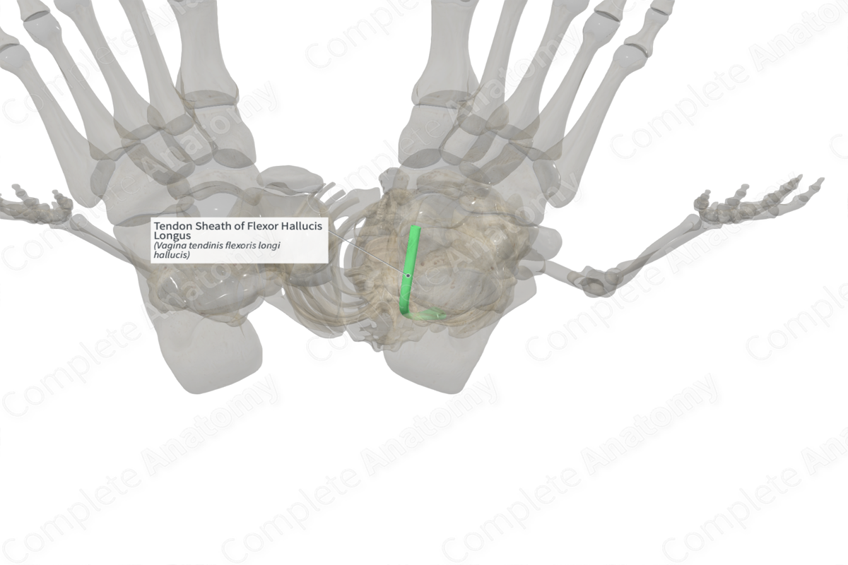
Tendon Sheath of Flexor Hallucis Longus (Right)
Vagina tendinis flexoris longi hallucis
Read moreStructure
Several tendons originating from muscles in the leg traverse the ankle to enter the foot. They pass beneath retinacula which holds the tendons in place, preventing subluxation. These tendons are covered by modified bursae, called tendon sheaths, which minimize friction with the surrounding structures. The sheaths, composed of both fibrous and synovial layers, wrap around the tendons, ensuring that the tendons can glide over adjacent structures and allow for minimal friction during movement.


Related parts of the anatomy
Anatomical Relations
The tendon sheath of the flexor hallucis longus muscle covers the tendons of this muscle as it travels deep to the flexor retinaculum of the foot, along the medial aspect of the ankle.
Function
The tendon sheath ensures that the tendons of the flexor hallucis longus muscle can glide over adjacent structures and allow for minimal friction during movement. Additionally, the tendon sheath produces synovial fluid, ensuring that the tendon is well lubricated.
Learn more about this topic from other Elsevier products
Tendon Sheath

Tenosynovectomy refers to the excision of thickened tendon sheath and other tissue surrounding a tendon, commonly seen in infection, chemical irritation, and rheumatoid arthritis (synovectomy).
