Anatomical Relations
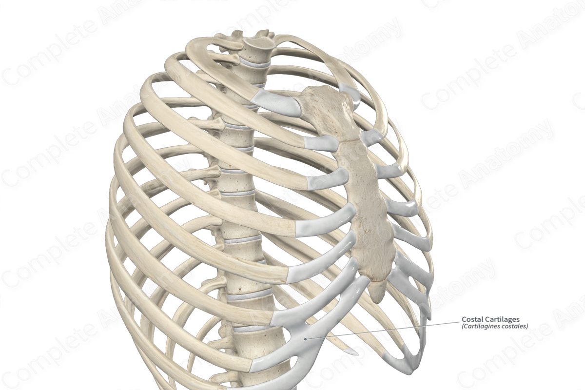
The costal cartilages form the anterior continuations of the ribs. The upper seven pairs join the sternum directly; the eighth, ninth, and tenth costal cartilages articulate with the inferior border of the cartilage above; the eleventh and twelfth cartilages have free ends that cover the tips of the eleventh and twelfth ribs.
The costal cartilages have:
—two surfaces, anterior and posterior;
—two borders, superior and inferior;
—two ends, medial and lateral.
The anterior surface of the costal cartilages is convex and faces anterosuperiorly. The costoclavicular ligament and subclavius muscle attach to the anterior surface of the first costal cartilages. Pectoralis major muscle attaches to the medial aspect of the anterior surfaces of the upper six cartilages, while the lower cartilages are covered by the superior attachments of the muscles of the anterior abdominal wall.
The posterior surface of the costal cartilages is concave and faces posteroinferiorly. The sternothyroid muscle attaches to the posterior surface of the first costal cartilages, transversus thoracic muscle attaches to the second to sixth cartilages, and transversus abdominis muscle attaches to the inferior six costal cartilages.
The external intercostal membranes and the internal intercostal muscles attach to the superior and inferior bonders of the costal cartilages, which are concave and convex, respectively.
The lateral end of each costal cartilage is continuous with its corresponding rib. The medial end of the first costal cartilage is continuous with the manubrium of the sternum, forming a fibrous synarthrosis (or suture) (Standring, 2016). The medial ends of the second to seventh costal cartilages are also continuous with the sternum; however, their ends are rounded and articulate with the costal notches on the lateral border of the sternum. These are synovial joints. The medial ends of the eighth, ninth, and tenth cartilages connect with the cartilages directly above them, while the medial borders of the eleventh and twelfth ribs are free.


Related parts of the anatomy
Structure
The costal cartilages are composed of hyaline cartilage that extends from the anterior ends of the ribs to the lateral surface of the sternum.
Function
Since the costal cartilages are composed of hyaline cartilage, they contribute to the mobility and elasticity of the thoracic cage, allowing for movement during respiration.
List of Clinical Correlates
—Forensic radiology (costal cartilage ossification)
References
Standring, S. (2016) Gray's Anatomy: The Anatomical Basis of Clinical Practice. Gray's Anatomy Series: Elsevier Limited.
