Anatomical Relations
The dorsal and palmar radioulnar ligaments are formed by the thickened dorsal and palmar aspects of the triangular fibrocartilaginous complex. The triangular fibrocartilaginous complex is both a cartilaginous and ligamentous structure that suspends the distal radius and medial carpal bones from the distal part of the ulna. It is composed of the articular disc of the radioulnar joint, the ulnocarpal meniscus, the ulnar collateral, dorsal and palmar radioulnar, palmar ulnocarpal ligaments, and the floor of the tendinous sheath of extensor carpi ulnaris (Standring, 2016).


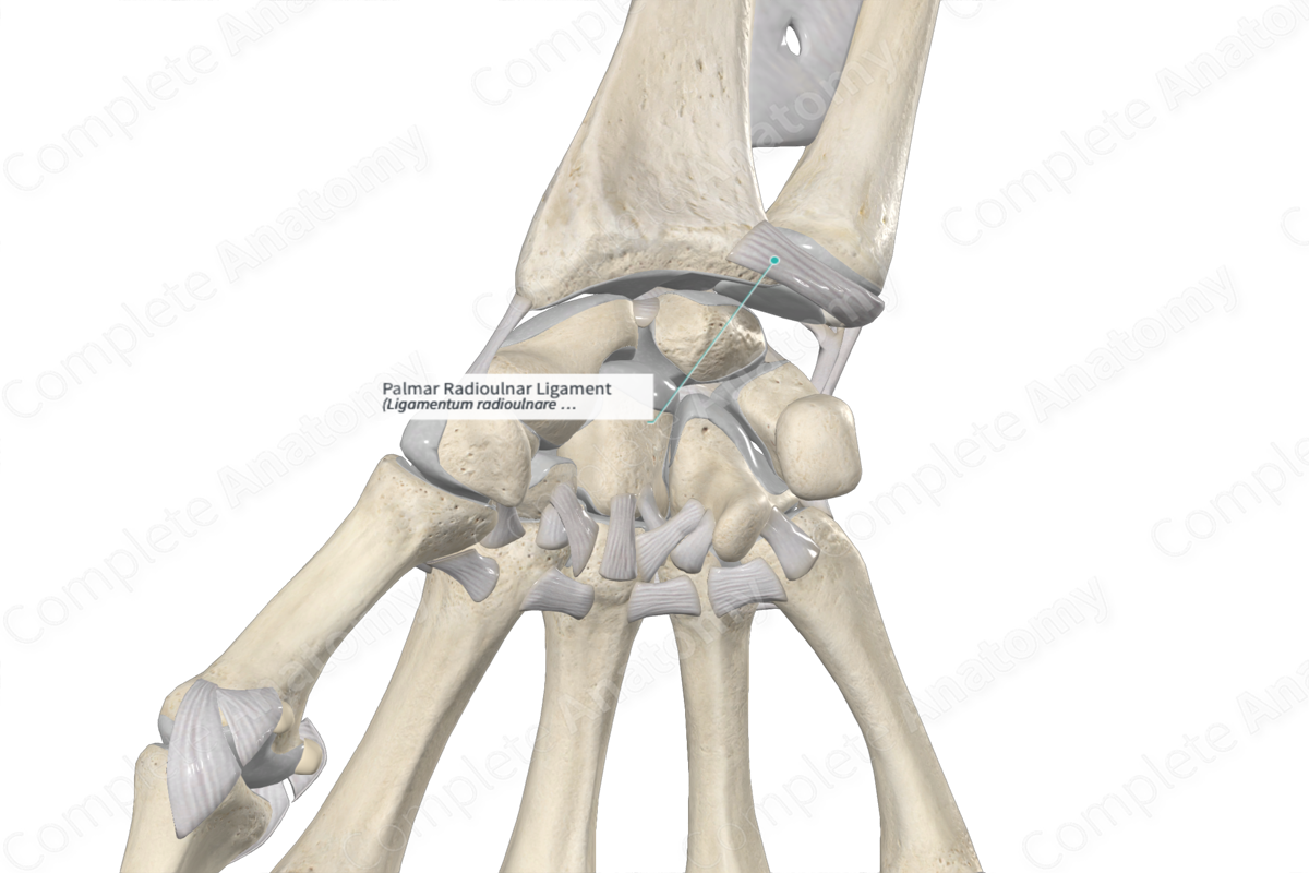
Structure
The palmar radioulnar ligament attaches to the palmar aspect of the distal radius and to the head of the ulna.
Function
The palmar radioulnar ligament forms part of the triangular fibrocartilaginous complex, which stabilizes the ulnocarpal and radioulnar joints, facilitates movements at the wrist joint, and transmits load from the carpal bones to the ulna.
References
Standring, S. (2016) Gray's Anatomy: The Anatomical Basis of Clinical Practice. Gray's Anatomy Series: Elsevier Limited.
Learn more about this topic from other Elsevier products

