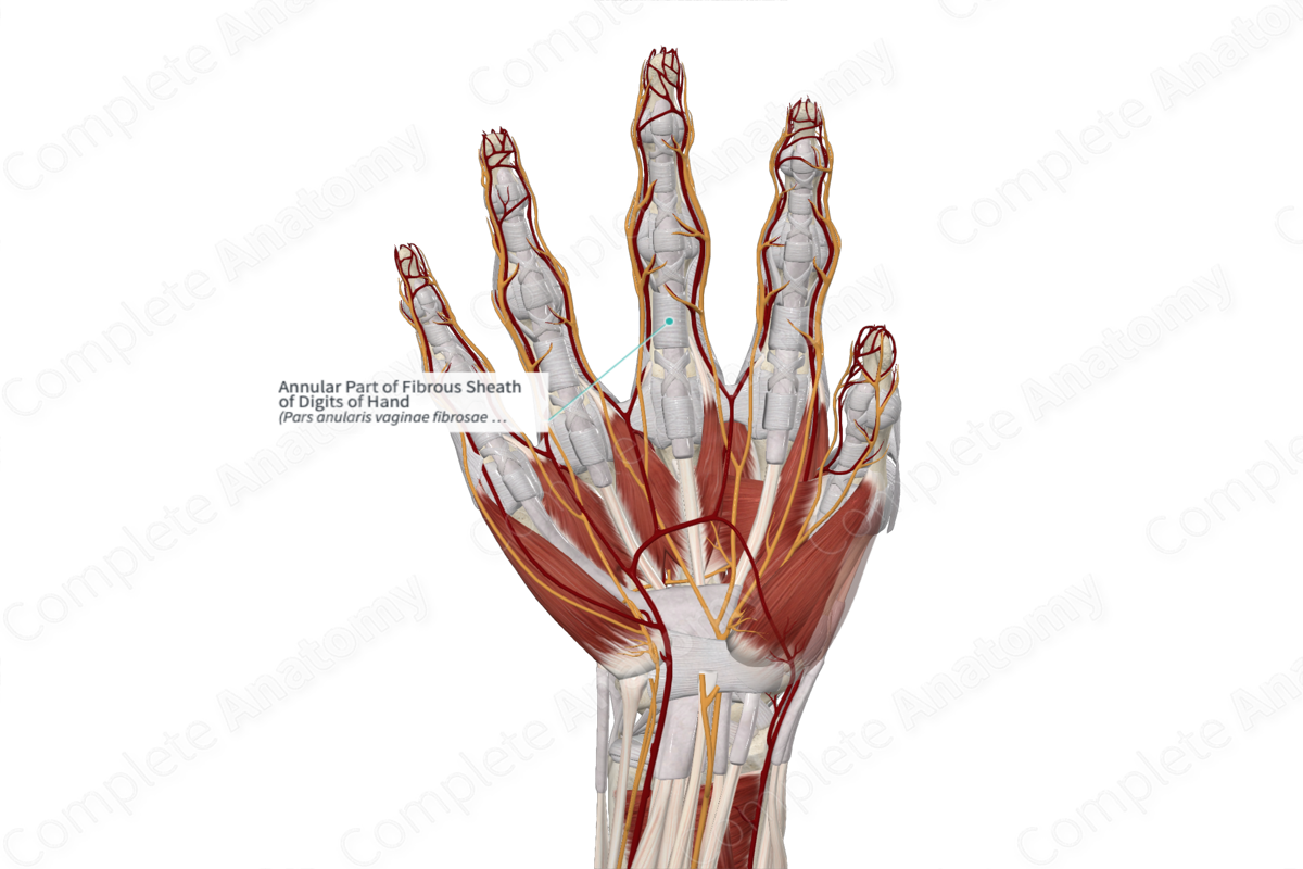
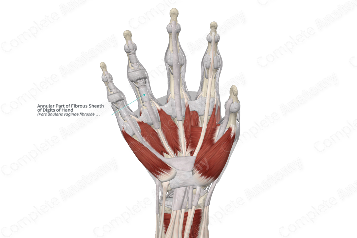
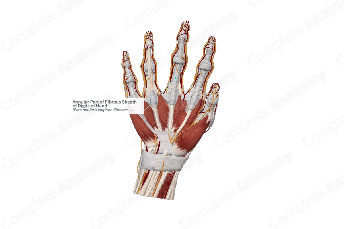
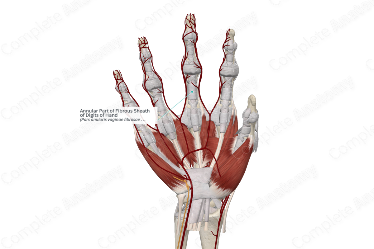
Annular Part of Fibrous Sheath of Digits of Hand
Pars anularis vaginae fibrosae digiti manus
Read moreAnatomical Relations
There are five regions of annular fibers named, from proximal to distal, A1—A5.
—A1 lies around the metacarpophalangeal joint and a portion of the proximal phalanx.
—A2 sits over the proximal phalanx and is the strongest portion of the pulley system.
—A3 is narrow and sits at the proximal interphalangeal joint.
—A4 sits on the mid region of the middle phalanx.
—A5 sits at the distal interphalangeal joint.
The most important pulley systems that prevent bowstringing are the A2 and A4 (Standring, 2016).
Structure
The word “annular” is the Latin for ring and thus annular fibers wind around the circumference of the phalanges of the digits of the hand.
Function
The annular part of the fibrous sheaths keep the tendons fixed in place on the bone and thus prevent bowstringing of the tendons.
References
Standring, S. (2016) Gray's Anatomy: The Anatomical Basis of Clinical Practice. Gray's Anatomy Series 41st edn.: Elsevier Limited.
Learn more about this topic from other Elsevier products

