Structure
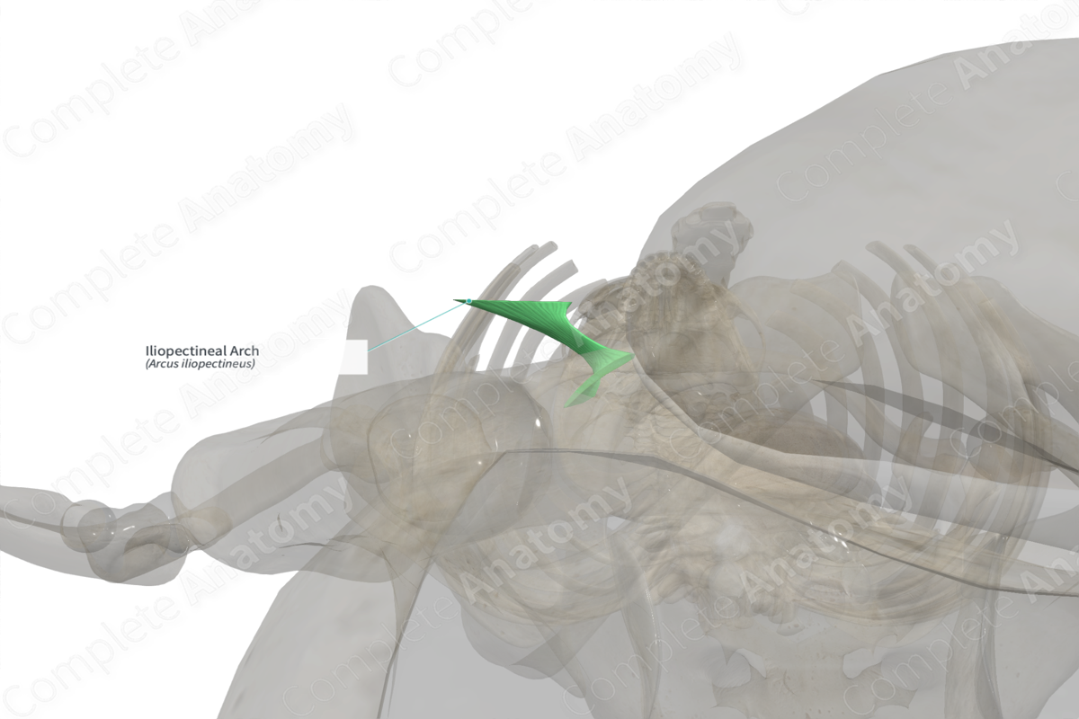
The iliopectineal arch is a condensed band of iliopsoas fascia. It is attached to the lateral third of the inguinal ligament and passes downwards and medially from the anterior superior iliac spine to the iliopectineal eminence of the pelvic bone. The iliopectineal arch divides the space beneath the inguinal ligament into muscular and vascular compartments.


Related parts of the anatomy
Anatomical Relations
The muscles of the anterior abdominal wall, internal abdominal oblique and transversus abdominis muscles, and the iliotibial tract arise from the iliopectineal arch. The iliopectineal arch crosses the lateral aspect of the femoral sheath. The femoral nerve runs below the iliopectineal arch on its inferior course.
Function
The iliopectineal arch forms a septum that divides the retro-inguinal space into the medial vascular compartment and the lateral muscular compartment. The femoral artery, femoral vein, lymphatics, and femoral canal run through the vascular compartment; while the femoral nerve and iliopsoas muscle run through the muscular compartment.
