Membranous Layer of Abdominal Subcutaneous Tissue
Stratum membranosum telae subcutanei abdominis
Read moreAnatomical Relations
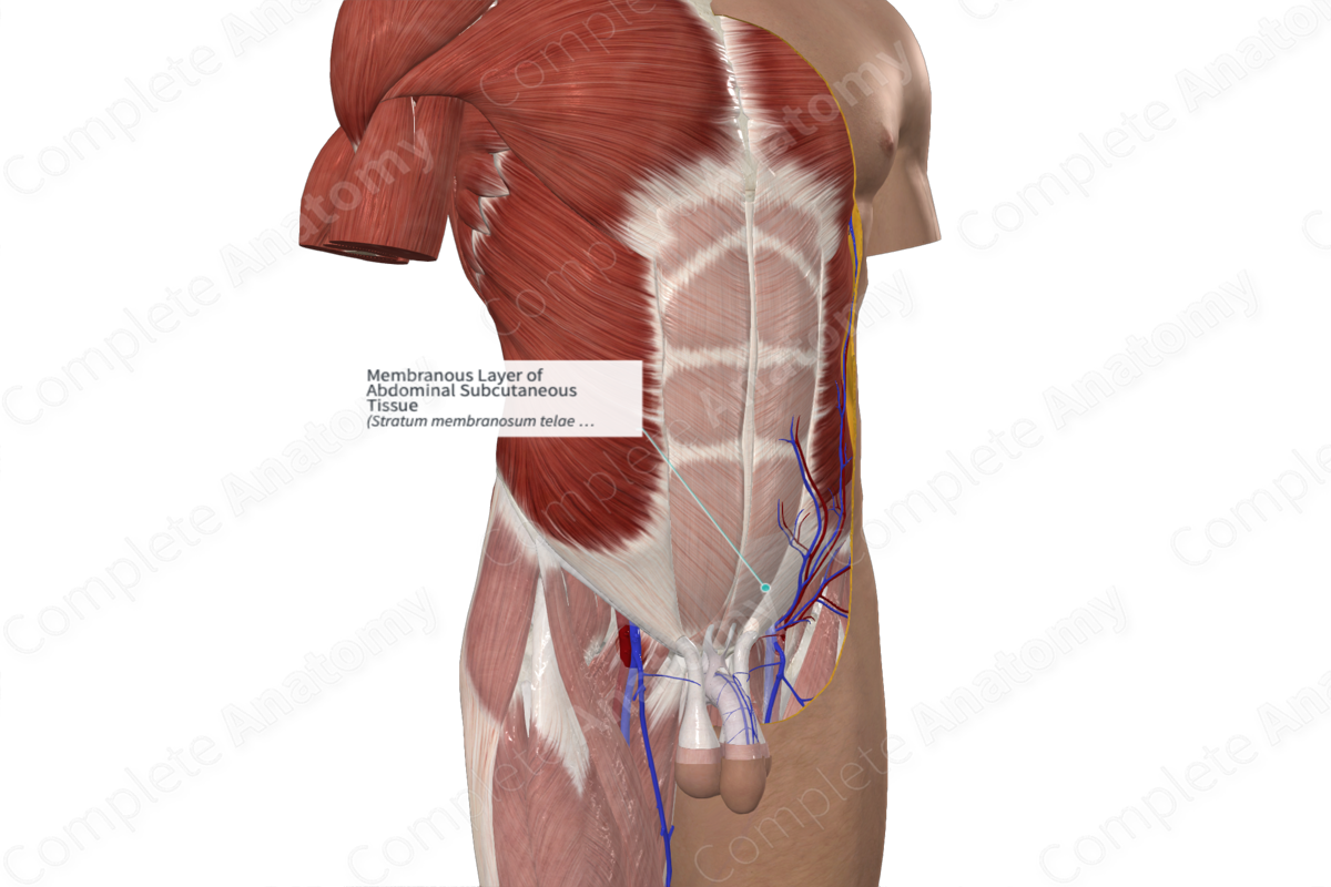
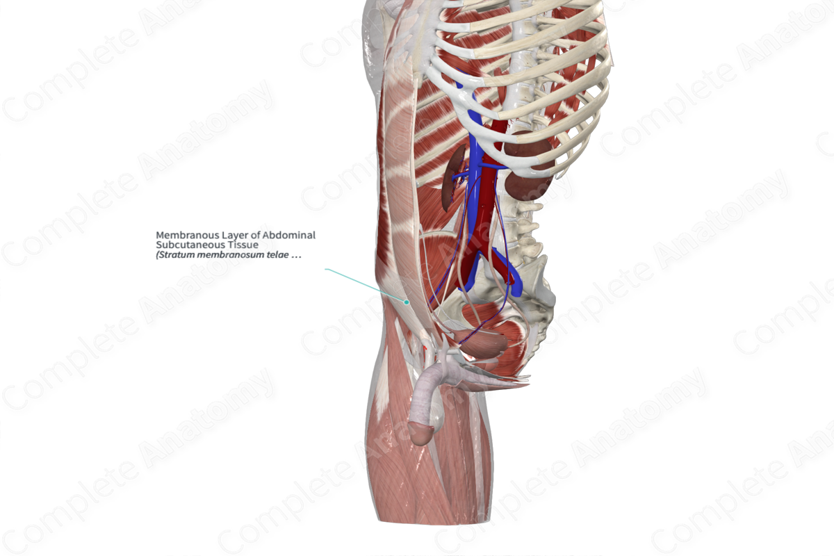
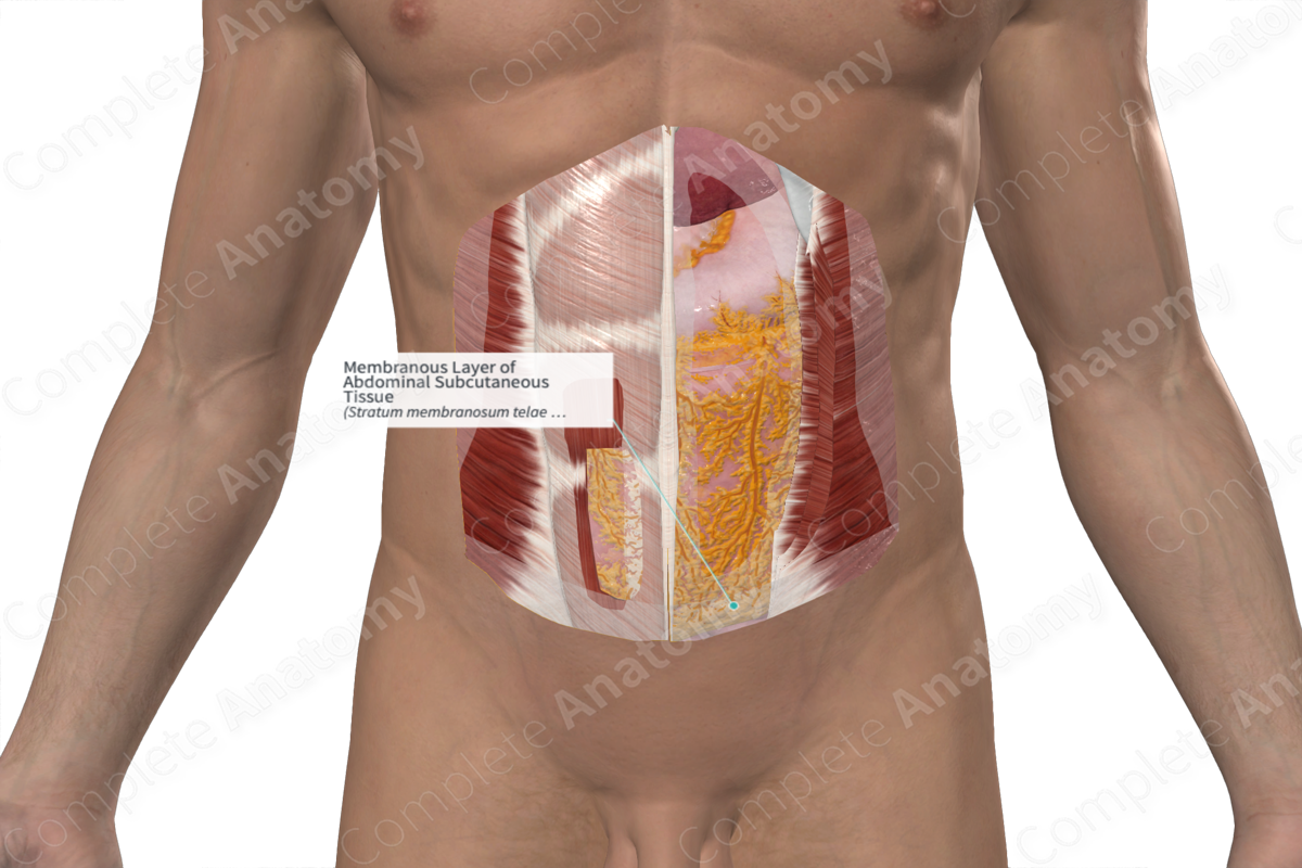
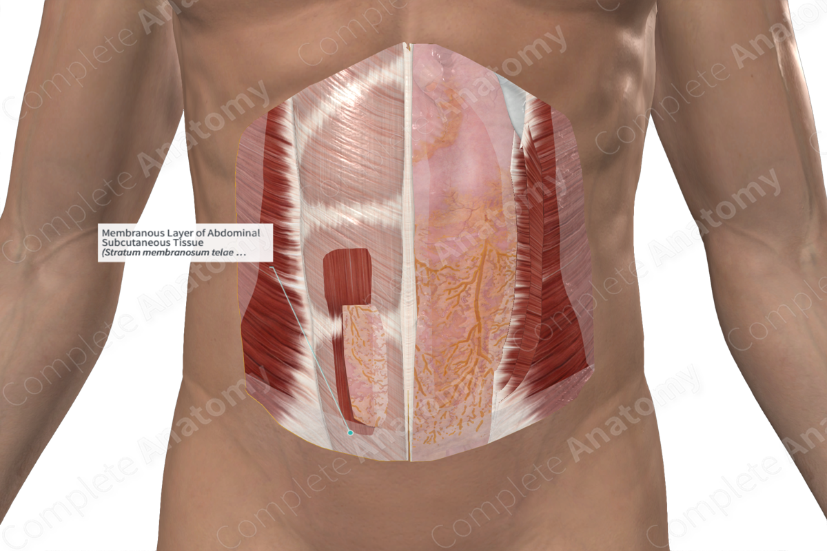
The membranous layer of abdominal subcutaneous tissue, or Scarpa’s fascia, is a thin membrane that is found under the skin and subcutaneous fat of the lower abdominal wall (Stecco and Hammer, 2014). Its upper attachment is at a point between the mid axillary line and the midclavicular line, along the costal margin, to a point approximately 2 cm below the umbilicus. It is continuous with the superficial fascia of the rest of the trunk superiorly. In the midline, Scarpa’s fascia is fused with the external abdominal oblique aponeurosis, rectus sheath, linea alba, pubic symphysis, and corresponding fascia on the opposite side. It blends with the deep fascia of external abdominal oblique muscle at the mid-axillary line and the thoracolumbar fascia posteriorly. Inferiorly, Scarpa’s fascia runs over the outer part of the inguinal ligament from the anterior superior iliac spine to the pubic tubercle, to fuse with the fascia lata of the thigh. It is continuous anteriorly and inferiorly with the perineal fascia (or superficial investing fascia of the perineum).
Structure
Scarpa’s fascia is a single, well-defined and continuous membrane. It is made up of fibrous tissue, elastic fibers, and adipose elements. It varies in size with an average of 0.5-1mm in thickness, 16–23 cm long, and 11–16 cm wide (Worseg et al., 1997). Scarpa’s fascia separates two distinct layers of fat; the deeper layer of loose areolar tissue separates it from the deep fascia of the anterior abdominal wall.
Function
The membranous layer of abdominal subcutaneous tissue plays an important role in the confining of fluid that has traveled from the retroperitoneum.
List of Clinical Correlates
—Eponymous clinical signs (Cullen’s sign, Grey Turner’s sign)
References
Stecco, C. and Hammer, W. I. (2014) Functional Atlas of the Human Fascial System E-Book. Elsevier Health Sciences.
Worseg, A. P., Kuzbari, R., Hübsch, P., Koncilia, H., Tairych, G., Alt, A., Tschabitscher, M. and Holle, J. (1997) 'Scarpa's fascia flap: anatomic studies and clinical application', Plast Reconstr Surg, 99(5), pp. 1368-80; discussion 1381.
Learn more about this topic from other Elsevier products


