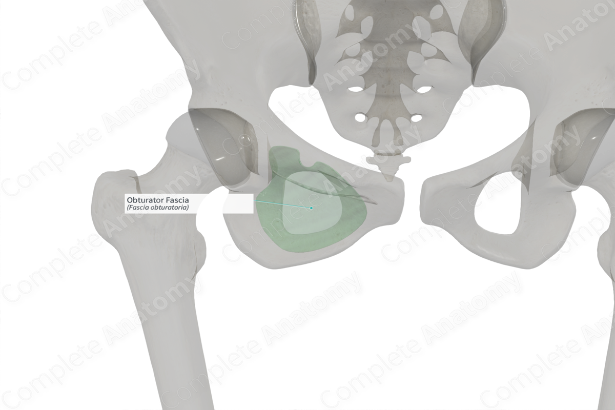
Anatomical Relations
The obturator fascia covers the pelvic surface of obturator internus muscle. It attaches to the posterior part of the pubis, the arcuate line of the ilium and is continuous with the piriformis fascia. Anteriorly and behind the pubis, it arches below the obturator vessels to give way for the obturator canal. The fasciae of the pelvic diaphragm/levator ani (mostly Iliococcygeus) and their corresponding muscles are attached to and suspended from a thickening of the obturator fascia called the tendinous arch of levator ani. The obturator fascia also has a horizontal passageway within it, called the pudendal canal; it lines the lateral wall of the ischioanal fossa and contains the pudendal artery, vein, and nerve as well as the nerve to the obturator internus. The obturator artery also runs anterolaterally on the obturator fascia.
Structure
The obturator fascia is thin above the tendinous arch of levator ani and forms the epimysium of the muscle. Anteriorly, it is continuous with the fasciae of the muscles of the deep perineal pouch.
Function
The medial surface of the obturator muscles and the overlying obturator fascia forms the lateral pelvic walls.
List of Clinical Correlates
—Pudendal Neuralgia
—Landmark for bladder repair for stress urinary incontinence
Learn more about this topic from other Elsevier products

