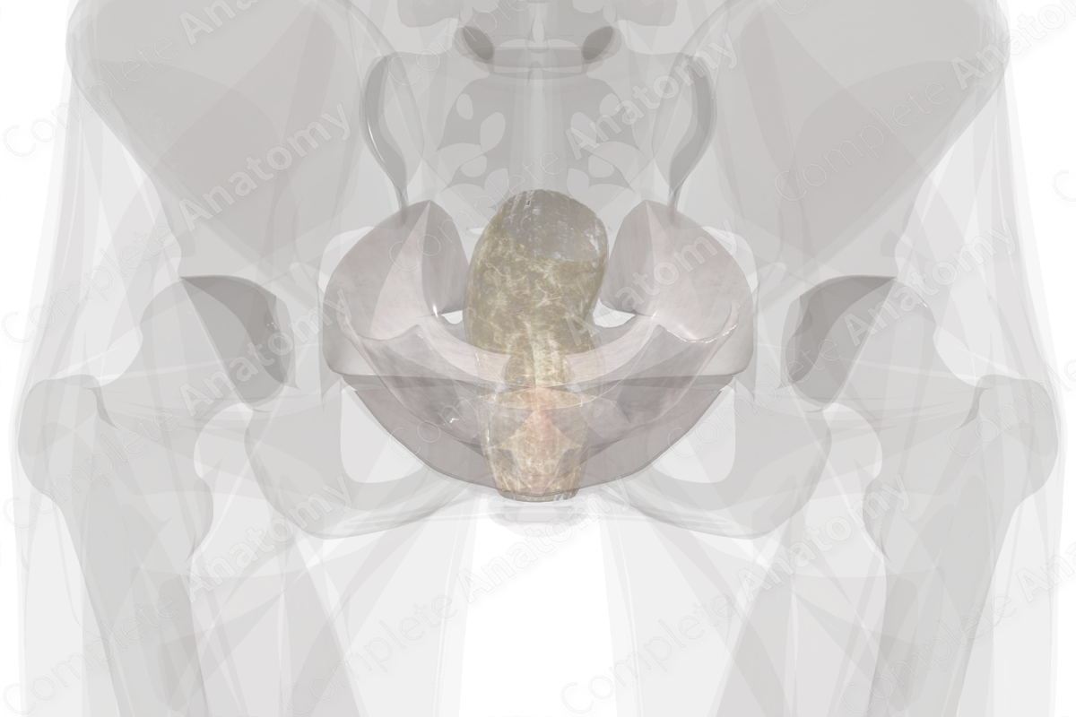
Anatomical Relations
The pelvic extraperitoneal fascia, or endopelvic fascia, sits inferior to the pelvic peritoneum and can be considered one continuous structure of connective tissue. It connects the urogenital organs, sitting within the midline of the pelvis, to the lateral pelvic walls. It connects to the pelvic wall primarily along the tendinous arch of pelvic fascia laterally, which runs from the pubic symphysis to the ischial spine. More posteriorly, it has a large expansion across the area of the piriformis muscles, which occupies the sciatic notch, sacroiliac joint, and sacrum (Standring, 2016).
This collection of loose connective tissue has areas of condensation, which are referred to as ligaments, such as the uterosacral and cardinal ligaments that adhere directly to the visceral fascia of the female urogenital organs. Endopelvic fascia related to the uterus is considered the parametrium and becomes the paracolpium as it continues inferior along the sides of the vaginal canal. The endopelvic fascia extends between the urogenital organs, separating them from one another and acting as packing material.
Posterosuperiorly, the endopelvic fascia extends a greater distance to reach the urogenital organs but anteroinferiorly, closer to the pubic symphysis and perineal membrane, the endopelvic fascia becomes much shorter.
Related parts of the anatomy
Structure
Endopelvic fascia is largely made up of loose connective tissue, which is easily dissected away from underlying structures. The areas of condensation referred to as ligaments, however, contain varying degrees of collagen and smooth muscle, hence their more fibrous appearance. Vasculature and nerves run throughout the endopelvic fascia.
Function
The endopelvic fascia carries out the role of supporting the pelvic urogenital organs and maintaining their position, while accommodating for changes in volume within each. The fascia also sheathes many neurovascular structures in its loose connective tissue as they travel through the pelvis.
List of Clinical Correlates
—Cystoceles
—Stress incontinence
—Genital organ prolapse
References
Standring, S. (2016) Gray's Anatomy: The Anatomical Basis of Clinical Practice. Gray's Anatomy Series 41st edn.: Elsevier Limited.


