Structure
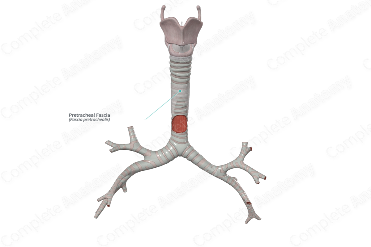
The pretracheal fascia is part of the visceral division of the deep cervical fascia. It lies anterior to the trachea.


Anatomical Relations
Superiorly, the pretracheal fascia is attached to the bottom of the hyoid bone. It runs inferiorly covering the anterior and lateral aspects of the trachea. Inferiorly, it is continuous with the fibrous pericardium. In the neck, it expands to cover the thyroid gland (Standring, 2016).
Function
The pretracheal fascia allows for frictionless movement of the component parts and anchors the thyroid gland during swallowing.
List of Clinical Correlates
—Myofascial pain
References
Standring, S. (2016) Gray's Anatomy: The Anatomical Basis of Clinical Practice, 41st ed. Elsevier Limited.
Learn more about this topic from other Elsevier products
Fascia

A fascia is a connective tissue that surrounds muscles, groups of muscles, blood vessels, and nerves.
