Structure
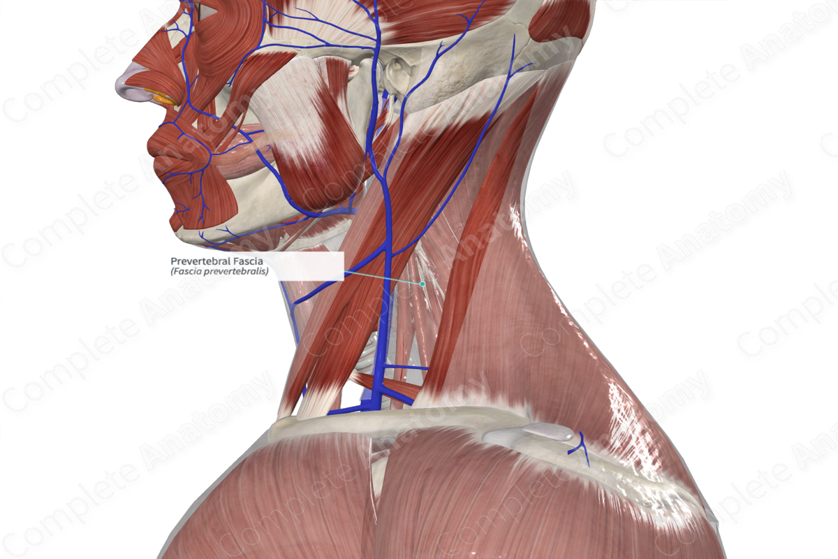
The prevertebral fascia is part of the deep investing cervical fascia. As the name suggests, is closely associated with the anterior aspect of the cervical vertebrae.
Related parts of the anatomy
Anatomical Relations
The prevertebral fascia extends from the base of the skull, superiorly, to the mediastinum, inferiorly, where it blends with the anterior longitudinal ligament as far down as T4 vertebra (varies between C6 to T4). It covers the anterior surfaces of the cervical vertebral bodies and the longus capitis and colli muscles and extends laterally and posteriorly to cover the scalene muscles, splenius capitis, and levator scapulae. As the subclavian artery and brachial plexus emerge between the scalenus anterior and medius, it drags a portion of the prevertebral fascia with it as the axillary sheath. Posteriorly, the prevertebral fascia attaches to the spinous processes of the cervical vertebrae (Standring, 2016).
Function
The prevertebral fascia ensheathes and protects the muscles and neurovasculature deep in the neck and allows fluid movements of the muscles over each other.
List of Clinical Correlates
—Compartment syndrome
—Endocarditis
—Mediastinitis
References
Standring, S. (2016) Gray's Anatomy: The Anatomical Basis of Clinical Practice, 41st ed. Elsevier Limited.
Learn more about this topic from other Elsevier products
Fascia

A fascia is a connective tissue that surrounds muscles, groups of muscles, blood vessels, and nerves.


