Transverse Intermuscular Septum of Leg
Septum intermusculare transversum cruris
Read moreStructure
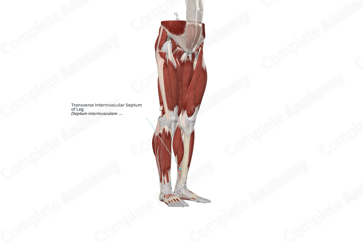
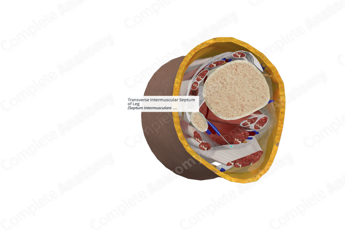
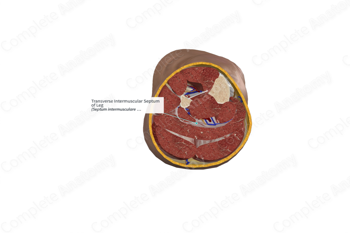
The transverse intermuscular septum is one of the three intermuscular septae of the lower leg, with the remaining two being the anterior and posterior intermuscular septae. These intermuscular septae are continuous with the crural fascia and separate the muscles of the leg into functional compartments.
The transverse intermuscular septum is thick and dense proximally, relatively thin in the middle leg, and thickens again distally (Standring, 2016). At its termination, it becomes reinforced with transverse fibers that extend between the tip of the medial malleolus and the calcaneus and, thus, forms the flexor retinaculum of the foot (Moore, Dalley and Agur, 2013).
Related parts of the anatomy
Anatomical Relations
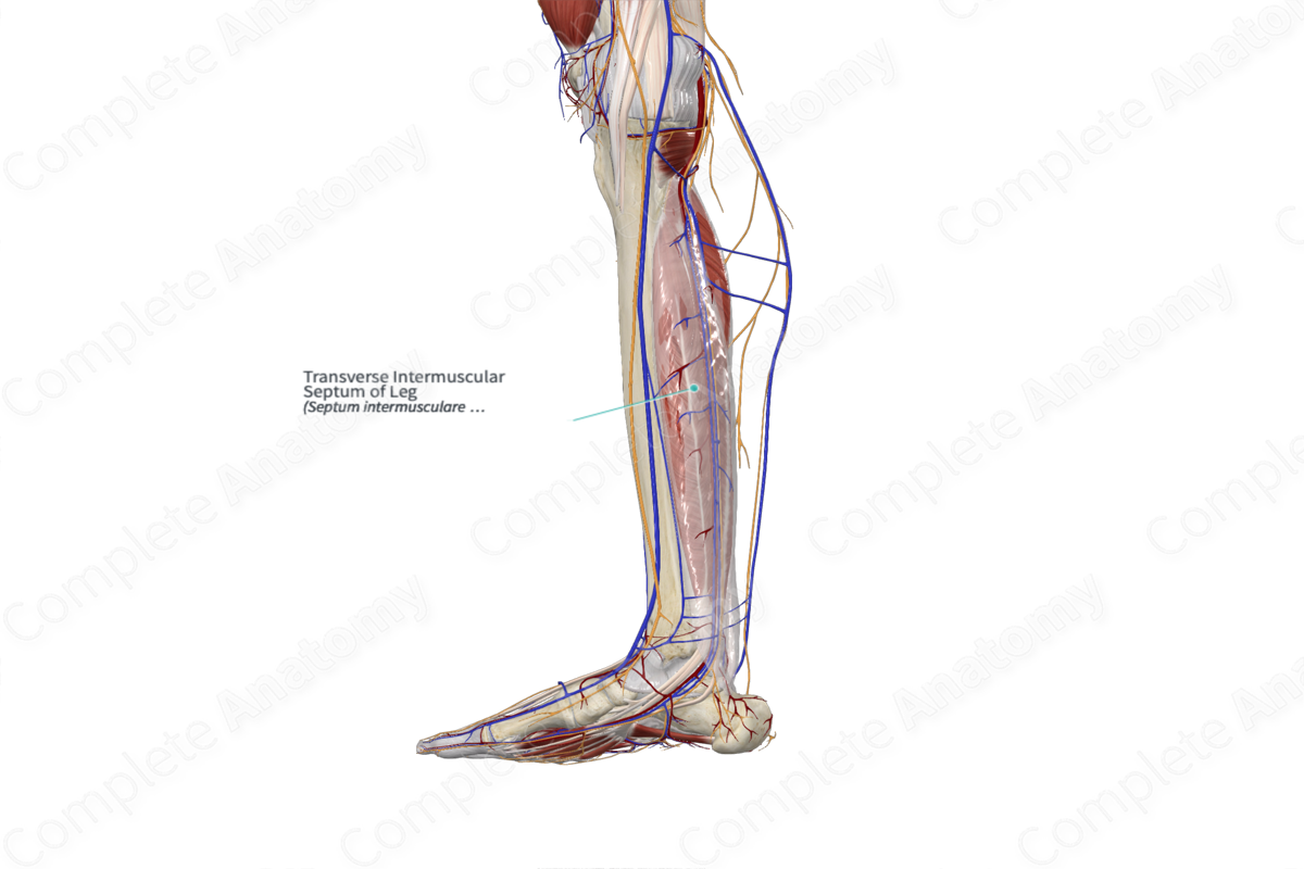
The transverse intermuscular septum is a fibrous stratum that lies between the superficial and deep muscles of the leg. It extends transversely from the medial margin of the tibia to the posterior border of the fibula. It separates the gastrocnemius and soleus muscles in the superficial part of the posterior compartment from the tibialis posterior, flexor hallucis longus, and flexor digitorum longus muscles in the deep part of the posterior compartment of the leg.
The transverse intermuscular septum is continuous distally with the flexor retinaculum of the foot and the superior fibular retinaculum. The posterior tibial artery passes deep to the transverse intermuscular septum (Standring, 2016).
Function
The transverse intermuscular septum divides the posterior compartment of the leg into superficial and deep parts.
List of Clinical Correlates
—Compartment syndrome
References
Moore, K. L., Dalley, A. F. and Agur, A. M. R. (2013) Clinically Oriented Anatomy. Clinically Oriented Anatomy 7th edn.: Wolters Kluwer Health/Lippincott Williams & Wilkins.
Standring, S. (2016) Gray's Anatomy: The Anatomical Basis of Clinical Practice. Gray's Anatomy Series 41st edn.: Elsevier Limited.


