Structure
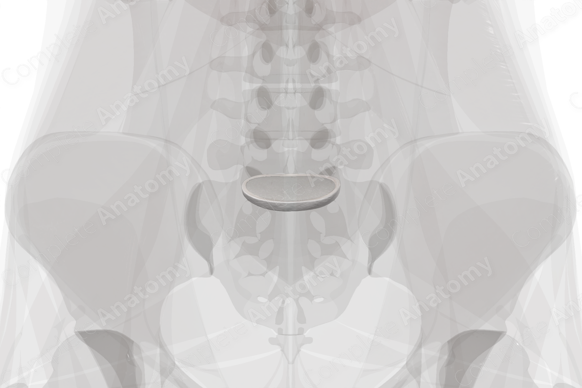
Symphyses are also known as secondary cartilaginous joints, i.e., they are made up of a fibrocartilage disc and the articular hyaline cartilages of two articulating bones. The lumbosacral symphysis is the cartilaginous joint that resembles the intervertebral symphyses. It is located between the vertebral body of the fifth lumbar with the body of the first sacral vertebra. The two vertebral bodies are interposed by a large intervertebral disc that is thickened anteriorly.
The lumbosacral symphysis is generally avascular but does receive nutrition by diffusion from vertebral bodies underneath the vertebral end plates. Venous drainage is via the external and internal vertebral venous plexuses.
The fifth lumbar vertebra and the sacrum also articulate at two zygapophyseal joints. Together, these joints and the lumbosacral symphysis are often collectively referred to as the lumbosacral junction.
Anatomical Relations
The lumbosacral symphysis is strengthened by the iliolumbar ligament.
Function
The lumbosacral symphysis transmits weight of the body through the sacrum and ilium, and on to the femur when standing, or the ischial tuberosities while seated. Additionally, flexion and extension occur at this joint.
Learn more about this topic from other Elsevier products


