Anatomical Relations
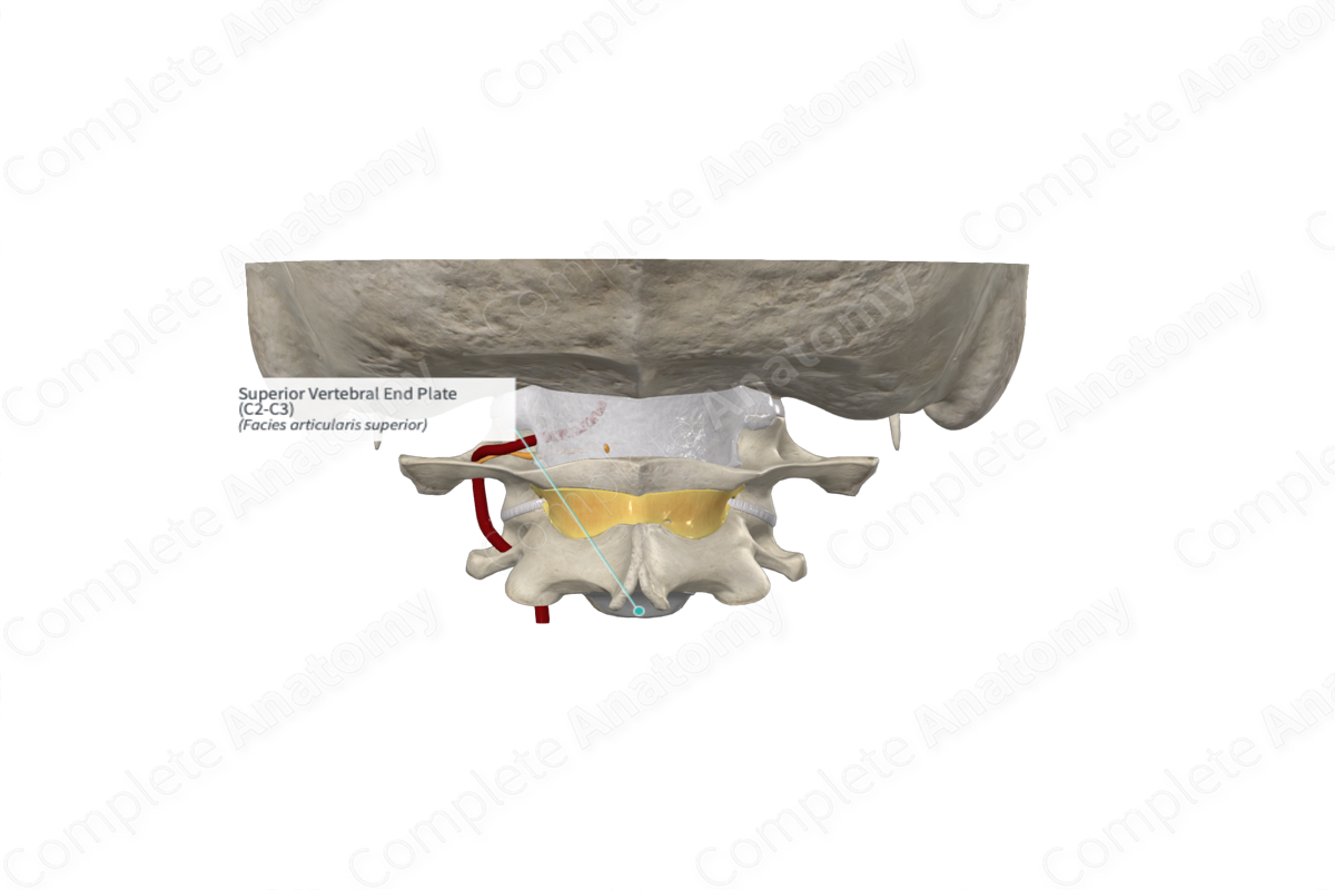
The superior vertebral end plate lies on the inferior surface of the vertebral body, which sits superiorly in the intervertebral joint. The end plates are surrounded by smooth bony epiphyseal rims, the annular epiphysis.


Related parts of the anatomy
Function
As a part of the intervertebral symphysis, the intervertebral end plates contribute to maintaining the stability of the vertebral column and resisting various forces in movement. They also help to maintain a fluid pressure of the water content of the nucleus pulposus, by preventing the loss of water to the vertebral body.
Structure
The superior vertebral end plates are bi-layered cartilages; they are flat and lie obliquely. End plates are relatively dense as they contain both hyaline cartilage and fibrocartilage. The fibrocartilage component lies nearer to the disc and is formed by the collagen fibers of the inner two thirds of the annulus fibrosus. The cartilage end plates are typically between 0.1-2.0 mm thick; however, this varies depending on the vertebral region and position (Lotz, Fields and Liebenberg, 2013). In the lumbar region, the end plates are thinner centrally than peripherally.
List of Clinical Correlates
—Disc Herniation
References
Lotz, J. C., Fields, A. J. and Liebenberg, E. C. (2013) 'The role of the vertebral end plate in low back pain', Global Spine J, 3(3), pp. 153-64.
