
Structure/Morphology
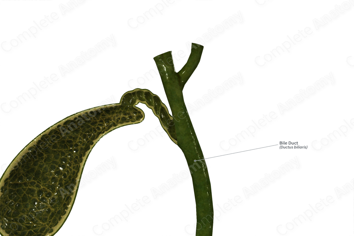
The bile duct is formed by the merging of the cystic and common hepatic ducts, and has three segments: supraduodenal, retroduodenal, and pancreatic.
Structurally, the bile duct has a mucosal layer and a muscular layer. The mucosal layer consists of a single cell epithelium on the luminal surface, small longitudinal folds, mucous glands, and loose connective tissue. The muscular layer consists of mostly connective tissue in the proximal portion of the duct, while the distal duct contains more smooth muscle fibers (Gulwani, 2012).


Key Features/Anatomical Relations
The bile duct runs up to 10 cm from its origin at the junction of the cystic and common hepatic ducts. It can be divided into three segments.
- The supraduodenal segment runs in the hepatoduodenal ligament to the right of the hepatic artery and to the right and anterior to the portal vein. It forms part of the portal triad.
- The retroduodenal segment runs posterior to the first part of the duodenum just right of the gastroduodenal artery.
- The pancreatic segment is typically embedded in the posterior surface of the head of the pancreas. It ends by the interface between pancreas and the second part of the duodenum. Here it joins the pancreatic duct to form the hepatopancreatic ampulla (of Vater), a small chamber deep to the major duodenal papilla (Standring, 2016).
Function
The bile duct transmits bile from either the liver or the gallbladder to the duodenum.
References
Gulwani, H. (2012) Histology-extrahepatic bile ducts. Gallbladder & extrahepatic bile ducts. https://www.pathologyoutlines.com/topic/gallbladdernormalhistologybileduct.html: PathologyOutlines.com (Accessed: August 7th 2020).
Standring, S. (2016) Gray's Anatomy: The Anatomical Basis of Clinical Practice. Gray's Anatomy Series 41 edn.: Elsevier Limited.
