Structure/Morphology
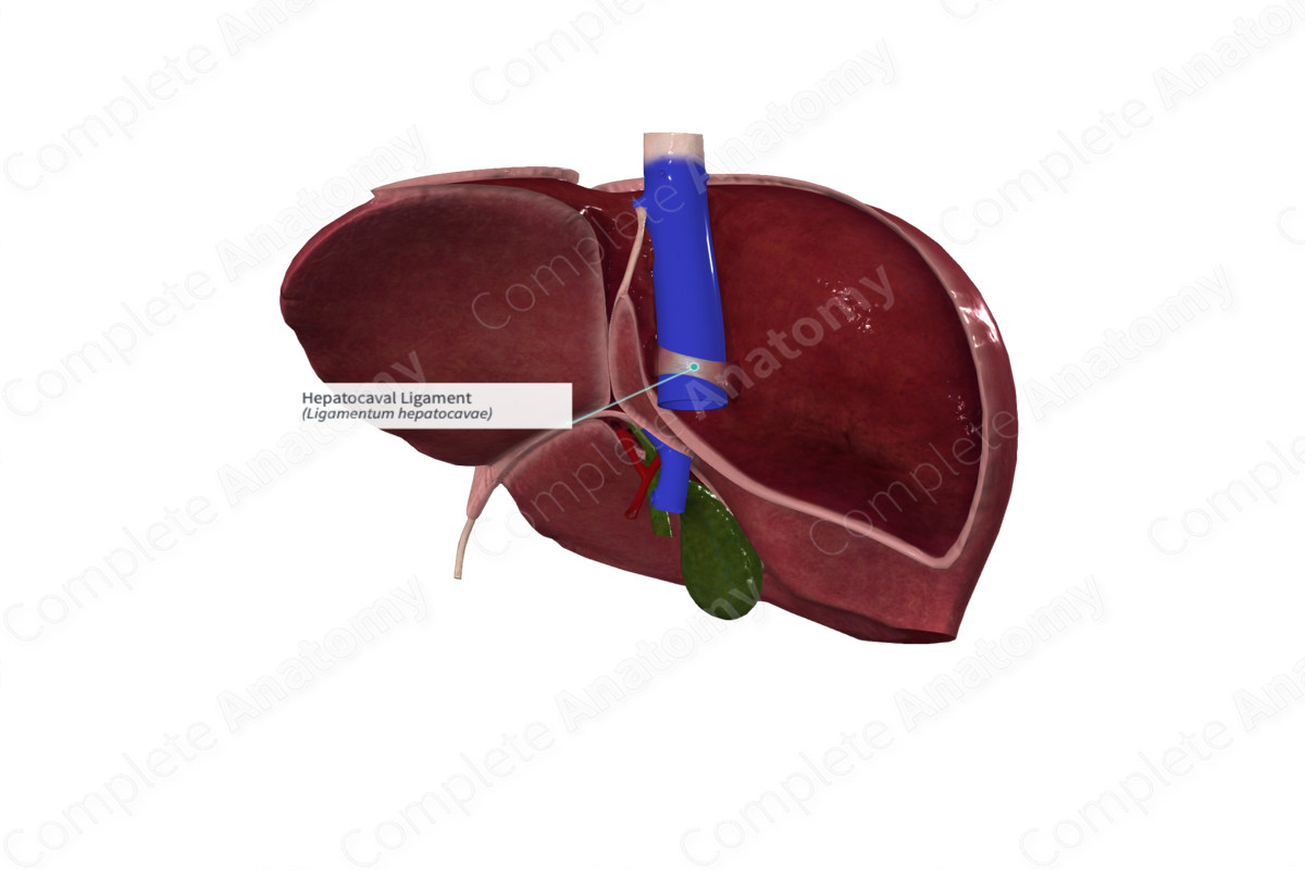
The hepatocaval ligament is the ligamentous remnant of liver parenchyma that enclosed the inferior vena cava posterior to the embryonic liver.


Key Features/Anatomical Relations
The hepatocaval ligament is found posterior to the inferior vena cava, anterior to the diaphragm, superior to the right suprarenal vein, and just to the left of the right suprarenal gland.
On the posterior surface of the inferior vena cava, it covers the inflow of the right hepatic vein. It stretches between segment VII of the right liver and segment I (or caudate lobe) of the left liver (Morjane et al., 2008).
List of Clinical Correlates
The hepatocaval ligament serves primarily as a landmark in surgical resections of the liver, particularly when the right hepatic vein must be accessed.
References
Morjane, A., Dahmane, R., Ravnik, D. and Hribernik, M. (2008) 'Anatomy and surgical relevance of the hepatocaval ligament. A study on cadaveric livers', Cells Tissues Organs, 187(3), pp. 243-6.
Learn more about this topic from other Elsevier products
Joint Ligament

Entheseal structures are widely located throughout the body and are represented by the interface between bone and several tissues including tendon, joint capsules and ligaments.