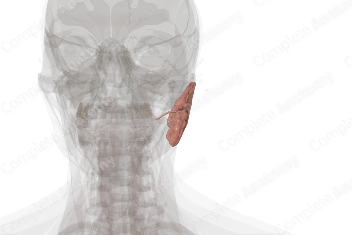
Parotid Gland (Left) Quick Facts
Location: Superficially in the lateral aspect of the face, anterior and inferior to the ear.
Arterial supply: External carotid artery.
Venous Drainage: External jugular vein.
Innervation: Sensory and parasympathetic innervation from the auriculotemporal nerve (CN V3).
Lymphatic drainage: Upper deep cervical lymph nodes.


Related parts of the anatomy
Parotid Gland (Left) Structure
The parotid gland is irregular in shape and consists of lobules of glandular tissue weighing approximately 25 g (Standring, 2016). The lobules are interspersed with adipose tissue and are covered in a layer of fascia, which forms the parotid capsule. This fascial layer derived from the deep cervical fascia of the neck.
Parotid Gland (Left) Anatomical Relations
The parotid gland extends from the posterior border of the ramus of the mandible to the upper portion of the sternocleidomastoid muscle in the neck. It sits anteroinferior to the external acoustic meatus and lies over the master muscle and mastoid process.
The parotid gland contains, from deep to superficial, the external carotid artery, the retromandibular vein, and the facial nerve with its five main branches (temporal, zygomatic, buccal, marginal mandibular, and cervical).
Although the gland is largely superficial, a smaller deep portion sits deep to the mandibular ramus on the lateral aspect of the superior constrictor muscle. Therefore, a deep lobe tumor may not present as a facial swelling, but as a protrusion in the lateral oropharyngeal wall.
Parotid Gland (Left) Function
The parotid gland secretes serous fluid upon parasympathetic stimulation. The influence of the sympathetic fibers is to reduce the secretions of the parotid gland.
Parotid Gland (Left) Arterial Supply
Branches arising from the external carotid artery supply the parotid gland. These include parotid branches of the posterior auricular and superficial temporal arteries, and the transverse facial artery.
Parotid Gland (Left) Venous Drainage
Venous drainage occurs via small unnamed branches draining into the external jugular vein.
Parotid Gland (Left) Innervation
The lesser petrosal nerve is a visceral motor nerve that supplies parasympathetic innervation to the parotid gland. It carries preganglionic parasympathetic fibers to the otic ganglion. There, the lesser petrosal nerve synapses with the postganglionic parasympathetic fibers that travel with the auriculotemporal nerve to innervate the parotid gland. The auriculotemporal nerve also conveys general sensory information from the parotid gland.
Parotid Gland (Left) Lymphatic Drainage
The parotid gland contains superficial and deep lymph nodes. Drainage of these nodes occurs through the deep lateral cervical nodes, primarily the internal jugular nodes either directly or via the infraauricular nodes. In 58% of cases, there is collateral drainage along lymph vessels that follow the course of the external jugular vein (Földi et al., 2012).
Parotid Gland (Left) List of Clinical Correlates
- Parotitis
- Facial paralysis
- Acute sialadenitis
- Parotid duct obstruction
- Xerostomia
Parotid Gland (Left) References
Földi, M., Földi, E., Strößenreuther, R. & Kubik, S. (2012) Földi's Textbook of Lymphology: for Physicians and Lymphedema TherapistsElsevier Health Sciences.
Standring, S. (2016) Gray's Anatomy: The Anatomical Basis of Clinical Practice. Gray's Anatomy Series 41 edn.: Elsevier Limited.
Learn more about this topic from other Elsevier products
Parotid Gland

The parotid space is defined as the parotid gland and the structures that are within it, such as the facial nerve, vessels, and the intraparotid lymph nodes.
