Structure/Morphology
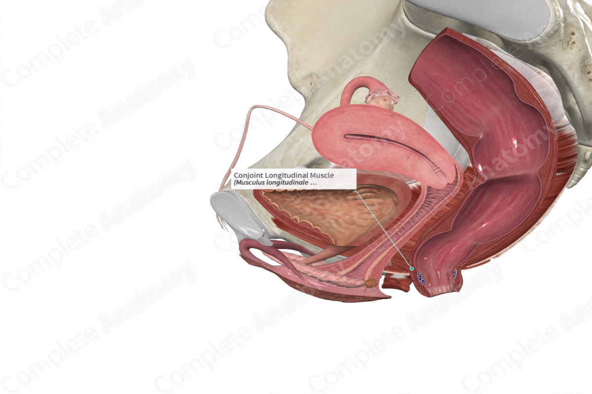
The conjoint longitudinal muscle is the continuation of the outer longitudinal muscle layer of the rectum as it traverses the anal canal (Standring, 2016).
Related parts of the anatomy
Key Features/Anatomical Relations
The conjoint longitudinal muscle sits between the internal anal sphincter and the external anal sphincter. Along the anal canal, its muscle fibers pass through the internal anal sphincter into the submucosal layer and aid in the formation of the submucosal muscle. Fibers also extend through the external anal sphincter and insert into the skin which aids in the formation of the puckered skin of the anal verge (Standring, 2016).
The conjoint longitudinal muscle layer is the most prominent during fetal development but with increased age it becomes increasingly fibrous (Haas, Fox and Haas, 1984).
Function
The conjoint longitudinal muscle is innervated by the autonomic nerves that supply the internal anal sphincter and contraction of this muscle is important during defecation. During this process this muscle helps to open the anal canal and flatten the anal cushions.
List of Clinical Correlates
- Hemorrhoids
References
Haas, P. A., Fox, T. A. and Haas, G. P. (1984) 'The pathogenesis of hemorrhoids', Dis Colon Rectum, 27(7), pp. 442-50.
Standring, S. (2016) Gray's Anatomy: The Anatomical Basis of Clinical Practice. Gray's Anatomy Series 41st edn.: Elsevier Limited.
