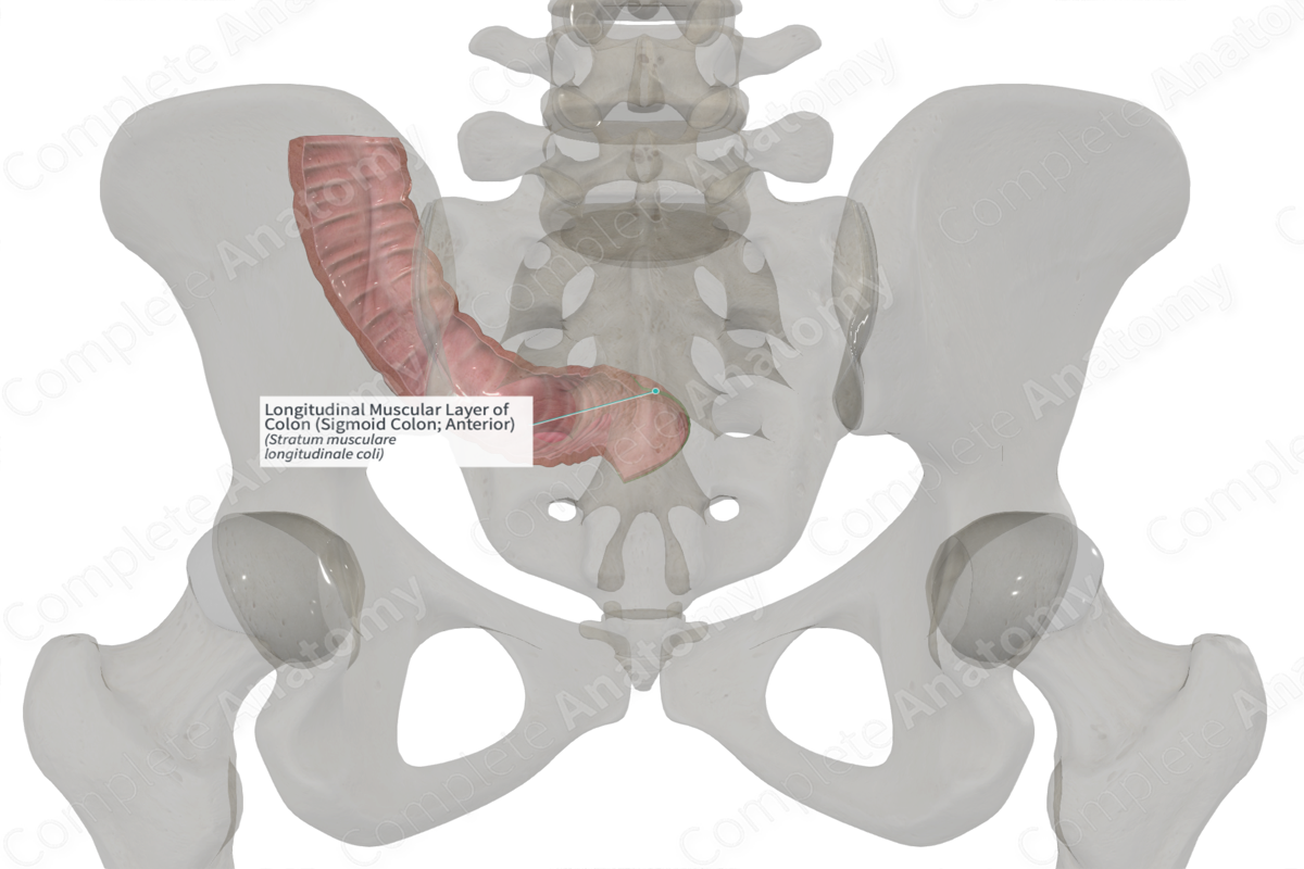
Longitudinal Muscular Layer of Colon (Sigmoid Colon; Anterior)
Stratum musculare longitudinale coli
Read moreLongitudinal Muscular Layer of Colon (Sigmoid Colon; Anterior) Structure/Morphology
The muscular layer of the colon, like the small intestine, consists of an inner circular layer and an outer longitudinal layer of smooth muscle. In the colon, the longitudinal layer is incomplete and forms three distinct bands, collectively termed the teniae coli (Standring, 2016).
Longitudinal Muscular Layer of Colon (Sigmoid Colon; Anterior) Key Features/Anatomical Relations
The three muscular bands sit beneath the outer serosal layer of the colon. They're named in relation to their position along the colon and are called the mesocolic, omental, and free teniae coli.
Longitudinal Muscular Layer of Colon (Sigmoid Colon; Anterior) Function
The longitudinal and circular layers of the muscular layer of the colon are responsible for producing peristaltic contractions of the gut, thus propelling the ingested substance through the lumen of the gut.
Longitudinal Muscular Layer of Colon (Sigmoid Colon; Anterior) References
Standring, S. (2016) Gray's Anatomy: The Anatomical Basis of Clinical Practice. Gray's Anatomy Series 41st edn.: Elsevier Limited.
Learn more about this topic from other Elsevier products
Colon

A faecaloma is a localized hard, inspissated, or calcified faecal mass, usually of a diameter equal to or greater than the colon.
