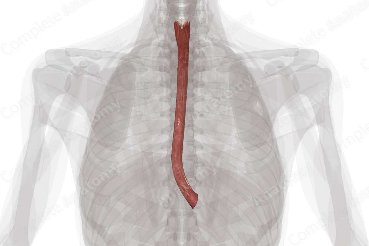
Longitudinal Muscular Layer of Esophagus
Stratum musculare longitudinale oesophagi
Read moreStructure/ Morphology
The circular and longitudinal muscular layers of the esophagus form the muscularis externa. The muscularis externa can be up to 300 μm thick and is composed of different types of muscle throughout its length. In the upper third, of the esophagus it is composed of skeletal muscle; the middle third is a mix of both skeletal and smooth muscle fibers; while the lower third is composed of smooth muscle only.
Related parts of the anatomy
Key Features/ Anatomical Relations
The longitudinal muscular layer is thicker than the circular layer and forms the outer layer of the muscularis externa. It extends the entire length of the esophagus, except posterosuperiorly, where it merges as two fascicles that ascend to the lower border of the inferior constrictor and ends as a tendon attaching to the posterior ridge of the cricoid lamina.
Function
The longitudinal layer of the muscular layer of the esophagus reduces the pressure force required to close the lumen of the esophagus, thus reducing the energy required by the circular layer of muscle during peristalsis (Brasseur et al., 2007).
References
Brasseur, J. G., Nicosia, M. A., Pal, A. and Miller, L. S. (2007) 'Function of longitudinal vs circular muscle fibers in esophageal peristalsis, deduced with mathematical modeling', World J Gastroenterol, 13(9), pp. 1335-46.


