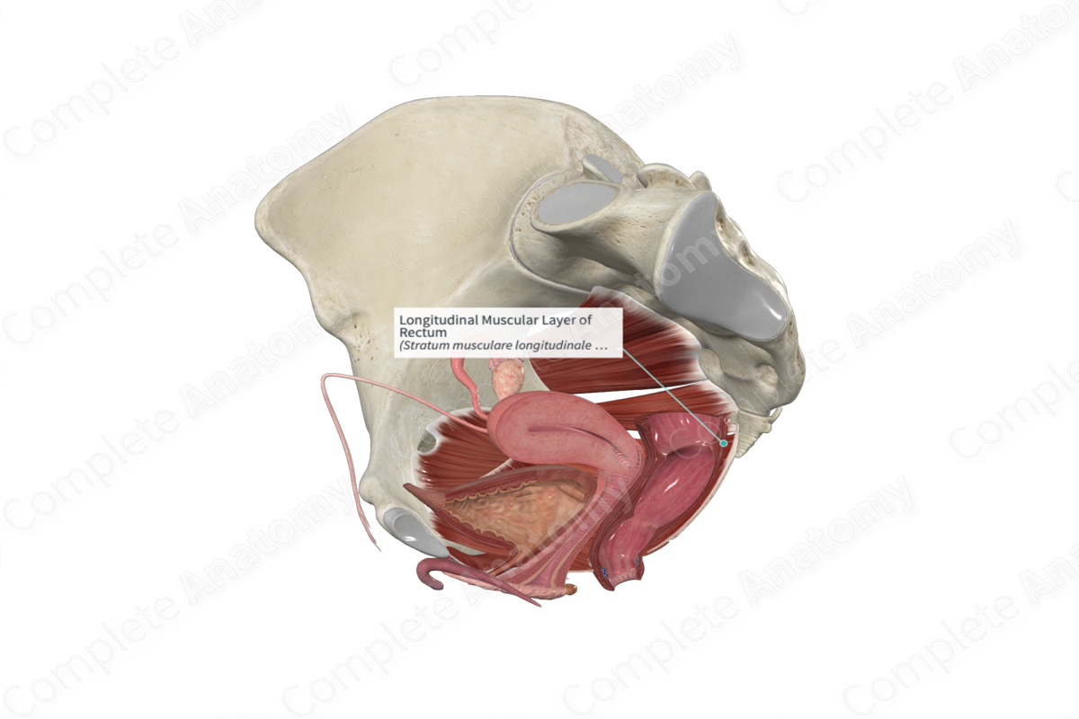
Longitudinal Muscular Layer of Rectum
Stratum musculare longitudinale recti
Read moreLongitudinal Muscular Layer of Rectum Structure/Morphology
The muscular layer of the rectum is composed exclusively of smooth muscle. Like the colon, it can be divided into inner and outer sublayers. The outer, longitudinal, sublayer has its muscle fibers oriented along the long axis of the rectum. Further, unlike the colon, the longitudinal layer of muscle, rather than being confined to three teniae coli spreads out to cover the entire surface of the rectum.


Longitudinal Muscular Layer of Rectum Key Features/Anatomical Relations
The lining of the rectum has two types of horizontal folds. The first type can be identified on the external surface and contains the mucosa, submucosa, the circular layer, and the longitudinal layers of muscle. The second type does not contain longitudinal muscle fibers and is only seen on the internal surface (Standring, 2016).
In males, some fibers of the longitudinal layer extend from the anterior rectal wall and insert into the perineal body to form the rectourethralis muscle.
Longitudinal Muscular Layer of Rectum Function
The longitudinal and circular layers of the muscular layer of the rectum are responsible for producing peristaltic contractions of the gut, thus propelling the ingested substance through the lumen of the gut.
Longitudinal Muscular Layer of Rectum References
Standring, S. (2016) Gray's Anatomy: The Anatomical Basis of Clinical Practice. Gray's Anatomy Series 41st edn.: Elsevier Limited.
Learn more about this topic from other Elsevier products
Rectum

The vasa recta are hairpin-shaped blood vessels that arise in the cortex and descend into the medulla before turning back into the cortex and leading to the venous system.
