Description
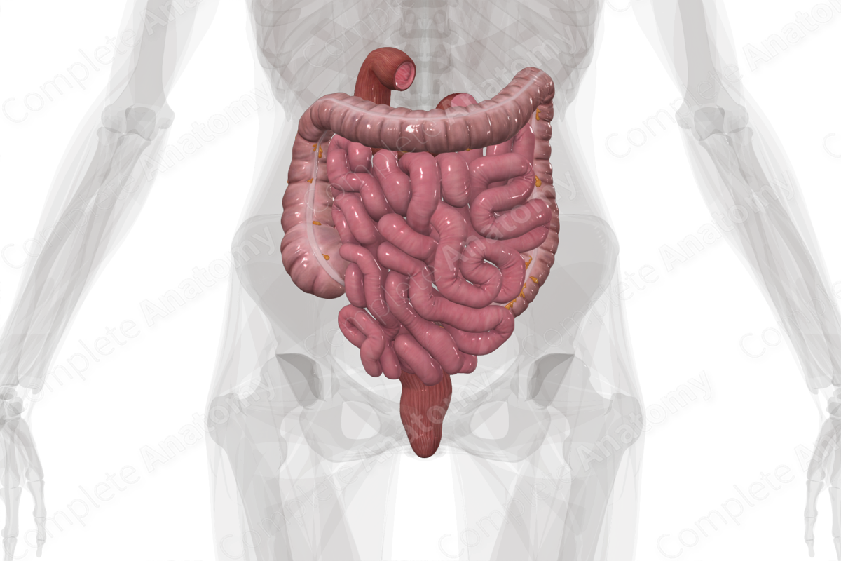
The digestive canal can be divided into upper and lower regions. The upper digestive canal includes the mouth, esophagus, and stomach. The lower digestive canal is formed by the small intestine (duodenum, jejunum, and ileum) and large intestine (cecum, colon, rectum, and anus).
Along its entire course, the digestive canal has a distinct four layered tubal wall. From inside out they include the: mucosa, submucosa, muscular layer, and serosa. Regional variations occur according to the development and function of each region.
Functionally, the lower digestive canal is responsible for digestion of chyme from the stomach, absorption of nutrients into the bloodstream, reabsorption of water and vitamins, and the solidification and excretion of wastes.
In the duodenum, acidic chyme is mixed with bile and pancreatic juices for further digestion. Bile, produced in the liver and stored in the gallbladder, is passed down the biliary ducts (hepatic, cystic, and common bile) and released into the duodenum through the hepatopancreatic ampulla (biliaropancreatic ampulla or ampulla of Vater). Joining it at this ampulla, the pancreatic duct carries enzymes synthesized in the pancreas (pancreatic juice). Bile neutralizes the highly acidic chyme and aids in the breakdown of lipids. Pancreatic enzymes act on the proteins, lipids, and carbohydrates to break them down further in preparation for absorption.
From the duodenum, chyme passes successively into the jejunum and ileum, subdivisions of the small intestine. As they move along their responsibility shifts from digestive to absorptive, so that eventually they primarily function to absorb essential nutrients, vitamins, bile salts and other small molecules. The ileum also plays host to an integral part in the immune system. Aggregations of lymphoid nodules called Peyer's patches are found throughout the ileum and, along with other mucosae associated lymphatic tissue (MALT), are responsible for combatting pathogenic microorganisms that may be ingested (Standring, 2016).
At the distal end of the ileum, what remains of the chyme, mostly waste products and water, is passed through the ileocecal valve into the cecum, the most proximal subdivision of the large intestine. The large intestines are responsible for reabsorbing water, salts, and vitamins from ileal wastes before final excretion.
Peristaltic contraction of the muscle walls lining the proceeding colonic segments push the ileal waste through the colon, where water reabsorption occurs, and thus solidifies the waste material. The colon consists of four segments, from proximal to distal: the ascending colon, the transverse colon, the descending colon, and the sigmoid colon.
At the terminal portion of the large intestine, compacted waste, now called feces or fecal matter, is stored in a highly distensible structure called the rectum. When the rectum fills, peristaltic contraction of the muscles lining its wall push the feces through the anal canal towards the anus. Opening of the anus is controlled by two muscles, the internal and the external anal sphincters. Control of the internal anal sphincter muscle is involuntary, while the external anal sphincter muscle can be controlled consciously to control the time and place of defecation.


Related parts of the anatomy
References
Standring, S. (2016) Gray's Anatomy: The Anatomical Basis of Clinical Practice. Gray's Anatomy Series 41st edn.: Elsevier Limited.
