Structure/ Morphology
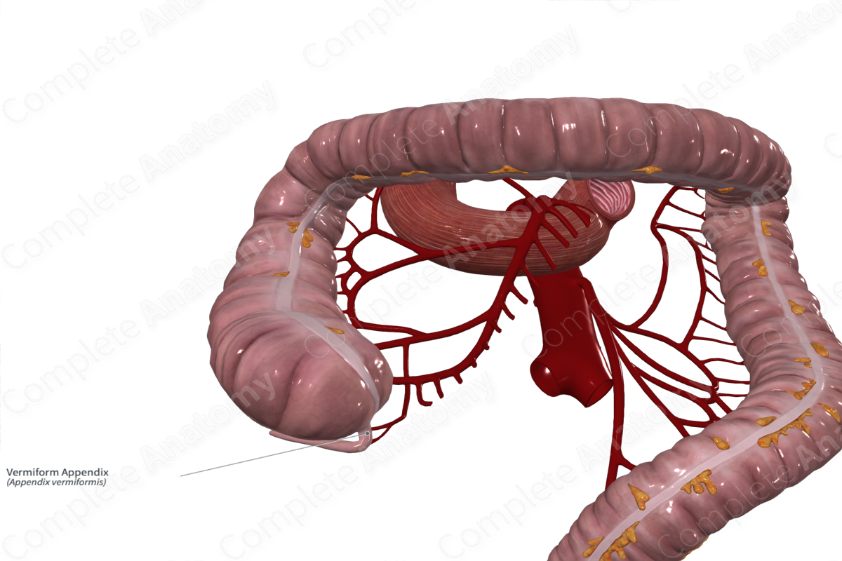
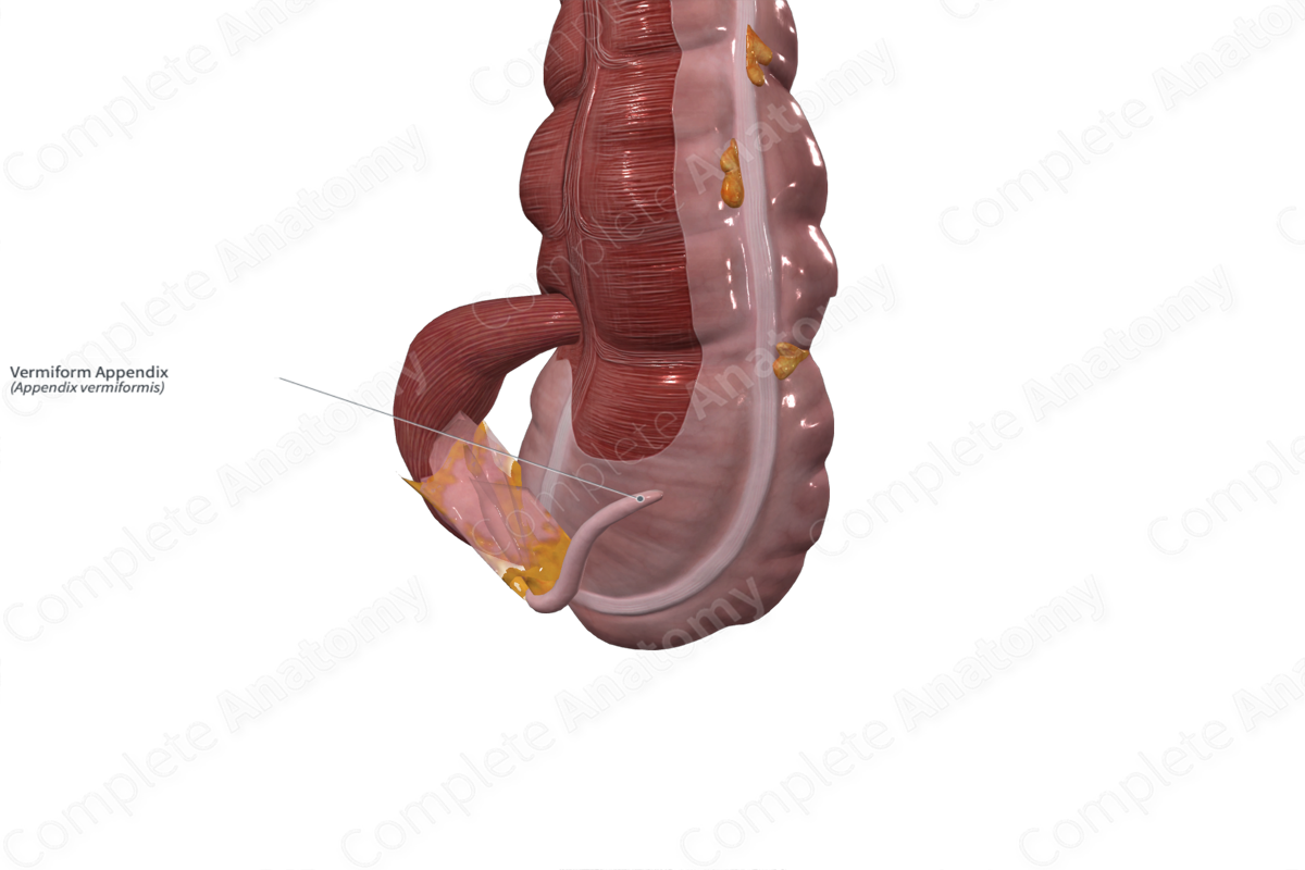
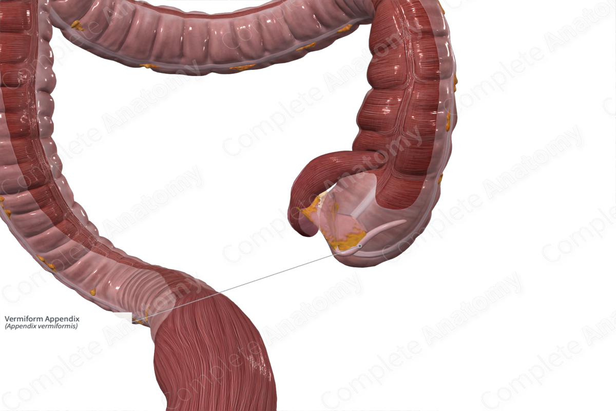
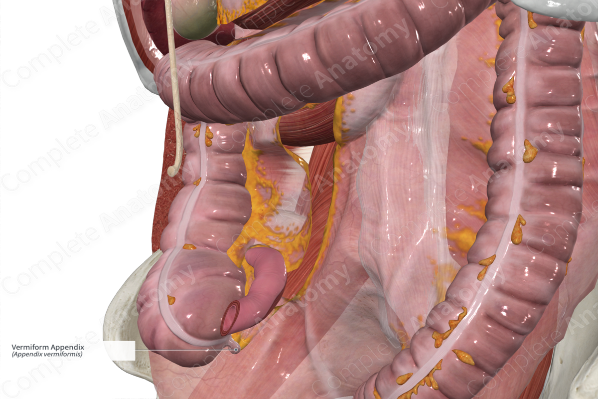
The appendix is a dead-end appendage that projects out from the posterior cecum below the ileocolic junction.
Significantly narrower than the large intestine, the appendix contains the same layers as the rest of the large intestine with some unique variations. The teniae coli are largely contiguous, forming a cohesive longitudinal muscle layer. Internally, the lumen is often obliterated, and the mucosa is enriched in lymphoid tissue. Crypts are present but at much lower concentrations than elsewhere in the large intestine (Standring, 2016).
Related parts of the anatomy
Key Features/Anatomical Relations
The appendix projects from the lower portion of the cecum, inferior to the ileocolic junction. It typically projects inferiorly 5–10 cm but can project in nearly any direction.
McBurney’s point (a spot on the skin two thirds of the way down a line between the umbilicus and the right anterior superior iliac spine) is often used to mark the approximate location of the tip of the appendix (Standring, 2016).
Function
The appendix is a vestigial organ that no longer serves the evolutionary role of digestion it plays in herbivores. However, increasing evidence suggests it serves as a reservoir for beneficial symbiotic bacteria, enabling recolonization of the colon if the normal microbiota is lost (Standring, 2016).
List of Clinical Correlates
- Acute Appendicitis
References
Standring, S. (2016) Gray's Anatomy: The Anatomical Basis of Clinical Practice. Gray's Anatomy Series 41 edn.: Elsevier Limited.
Learn more about this topic from other Elsevier products


