Quick Facts
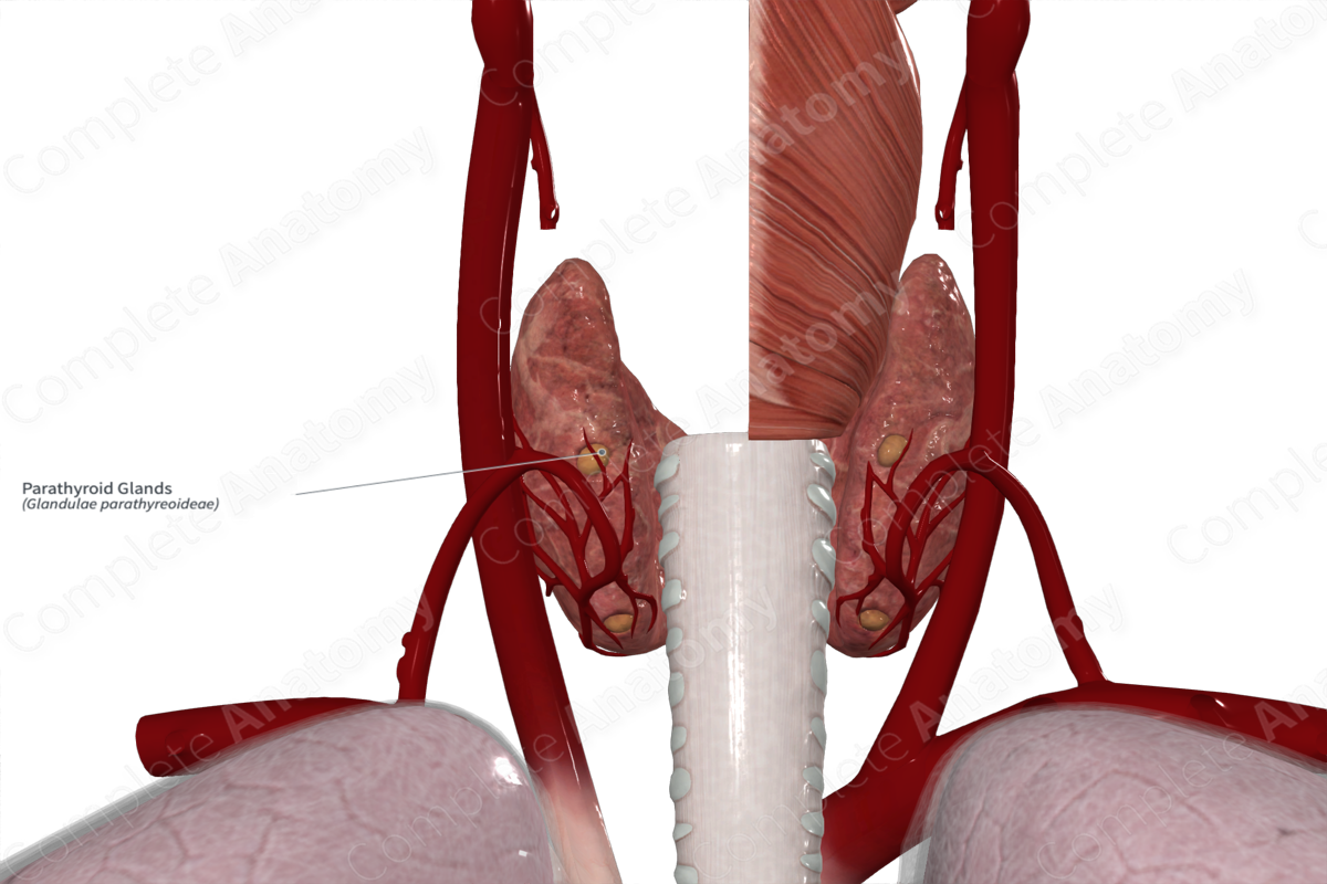
Location: Posterior surface of the thyroid gland.
Arterial Supply: Inferior thyroid artery.
Venous Drainage: Venous plexus along the anterior surface of the thyroid gland.
Innervation: Sympathetic innervation from the superior or middle cervical ganglion.
Lymphatic Drainage: Along the thyroid and thymus glands.


Related parts of the anatomy
Structure/Morphology
The parathyroid glands are paired ovoid structures lying on the posterior surface of the thyroid gland. There are two superior and two inferior glands that typically measure 6 mm long, 3 mm wide, and 1 mm from anterior to posterior. These glands are principally responsible for the production and release of parathyroid hormone (PTH) (Standring, 2020).
Anatomical Relations
The paired parathyroid glands reside along the posterior surface of the thyroid gland.
Function
The parathyroid glands are principally responsible with synthesizing and secreting parathyroid hormone (PTH), which directly relates to the levels of calcium and phosphorus in the blood. When blood calcium levels are decreasing, the production and release of PTH increases.
Arterial Supply
Both the superior and inferior parathyroid glands are supplied by the inferior thyroid artery. The superior glands are also occasionally supplied by the superior thyroid arteries.
Venous Drainage
The parathyroid glands are drained by a venous plexus on the anterior thyroid surface.
Innervation
The parathyroid glands receive sympathetic innervation from the superior or middle cervical ganglia (via thyroid branches).
Lymphatic Drainage
The cervical paratracheal lymph nodes receive afferent vessels from the thyroid and parathyroid glands. The efferent vessels of the cervical paratracheal lymph nodes pass to the internal jugular nodes of the deep lateral cervical lymph nodes (Földi et al., 2012).
List of Clinical Correlates
- Thyroidectomy
- Parathyroidectomy
- Hypoparathyroidism,
- Hyperparathyroidism
References
Földi, M., Földi, E., Strößenreuther, R. and Kubik, S. (2012) Földi's Textbook of Lymphology: for Physicians and Lymphedema Therapists. Elsevier Health Sciences.
Standring, S. (2020) Gray's Anatomy: The Anatomical Basis of Clinical Practice. 42nd edn.: Elsevier Health Sciences.
