Quick Facts
Location: Anterior to the larynx and trachea.
Arterial Supply: Superior and inferior thyroid arteries.
Venous Drainage: Superior, middle, and inferior thyroid veins.
Innervation: Superior, middle, and inferior cervical ganglia.
Lymphatic Drainage: Prelaryngeal or deep cervical nodes.


Related parts of the anatomy
Structure/Morphology
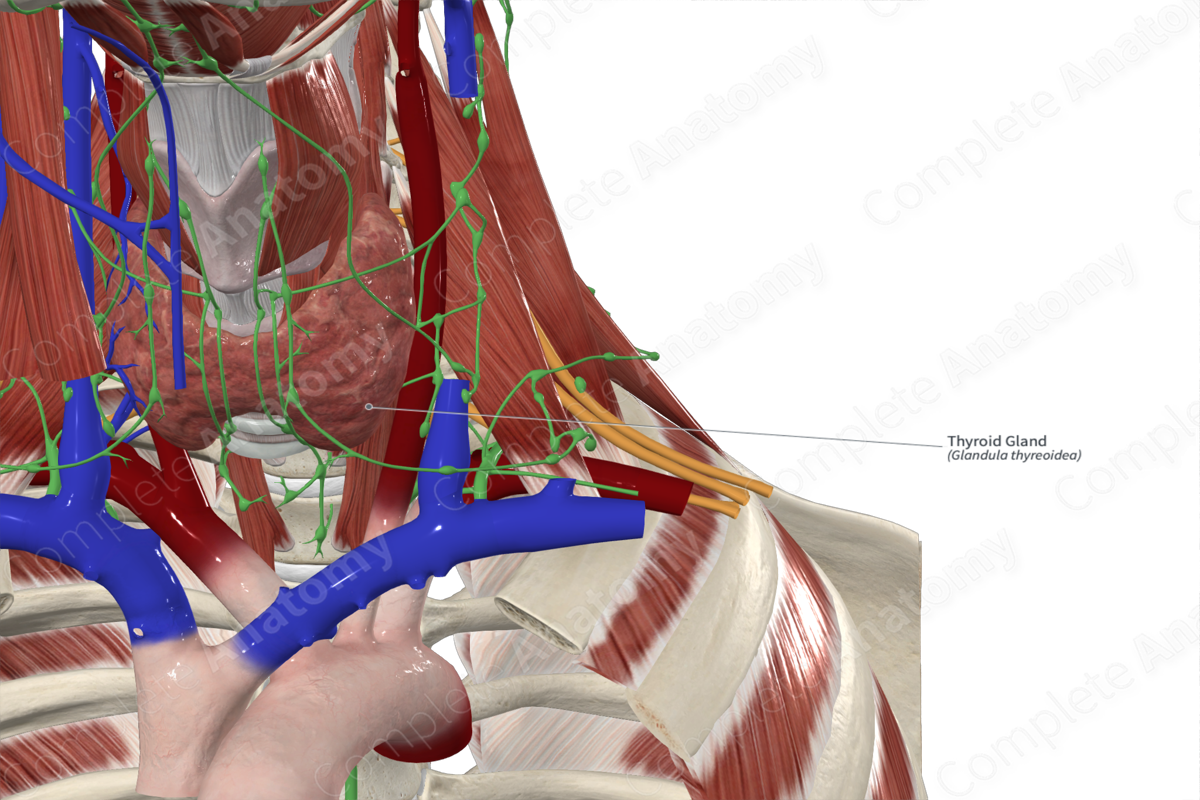
The thyroid gland is a large gland residing in anterior neck along the inferior larynx and superior trachea. The gland is one of the largest endocrine glands. It is divided into left and right lobes typically connected by the isthmus. Each lobe is approximately 5 cm long (Standring, 2020).
The thyroid gland has a rich blood supply and innervation and responsible for synthesizing and releasing two thyroid hormones. These hormones directly regulate important processes like metabolism and protein synthesis.
Anatomical Relations
The thyroid gland is covered by the sternothyroid, sternohyoid, and omohyoid muscles anteriorly and the sternocleidomastoid muscle inferiorly. The superior pole of the gland contacts the inferior pharyngeal constrictor muscle and the side of the cricoid cartilage.
Function
The thyroid gland synthesizes the thyroid hormones (triiodothyronine [T3] and thyroxine [T4]). These hormones are important for growth, cell differentiation, control of basal metabolic rate, and oxygen consumption in cells. These hormones also affect protein, lipid, and carbohydrate metabolism.
Arterial Supply
The superior and inferior thyroid arteries (and occasionally the thyroid ima artery) supplies the thyroid gland. These arteries anastomose with one another on the same side and on the contralateral side. The superior thyroid artery divides into an anterior branch to supply the anterior surface, and a posterior branch to supply the lateral and medial surfaces of the gland. The inferior thyroid artery divides into superior (ascending) and inferior branches, which supply the inferior and posterior surfaces of the gland. The superior branch also supplies the parathyroid glands.
Venous Drainage
Superior, middle, and inferior thyroid veins drain the thyroid gland. The superior thyroid vein drains the upper part of the gland and drains into the internal jugular vein. The middle thyroid vein drains the inferior part of the gland and drains into the internal jugular vein. The inferior thyroid veins arise from a thyroid venous plexus which also connects to the middle and superior thyroid veins. The left and right inferior thyroid veins drain into the left and right brachiocephalic veins respectively.
Innervation
The superior, middle, and inferior cervical ganglia principally innervate the thyroid gland. Postganglionic fibers from the inferior cervical ganglion forms a plexus along the inferior thyroid artery to travel to the gland.
Lymphatic Drainage
Lymph from the thyroid gland is drained by the prelaryngeal and thyroid nodes located above and below the isthmus. Their efferents carry lymph to the pre- and paratracheal nodes. Laterally, the gland is drained by the deep anterior cervical nodes. Some of the lymph vessels may also drain directly into the thoracic duct (Standring, 2020).
List of Clinical Correlates
- Thyroidectomy
- Parathyroidectomy
References
Standring, S. (2020) Gray's Anatomy: The Anatomical Basis of Clinical Practice. 42nd edn.: Elsevier Health Sciences.
Learn more about this topic from other Elsevier products

Thyroid and parathyroid gland histology: Video, Causes, & Meaning

Thyroid and parathyroid gland histology: Symptoms, Causes, Videos & Quizzes | Learn Fast for Better Retention!
