Quick Facts
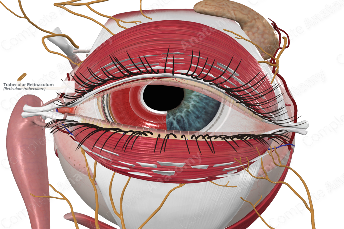
The trabecular retinaculum is a trabecula of loose fibers found at the iridocorneal angle between the anterior chamber of the eye and the venous sinus of the sclera; the aqueous humor filters through the spaces between the fibers into the sinus and passes into the bloodstream. The reticulum is divided into a corneoscleral part and a uveal part (Dorland, 2011).


Related parts of the anatomy
Structure and/or Key Feature(s)
The trabecular retinaculum, or meshwork, is a triangular-shaped region of connective tissue found at the intersection of the sclera, cornea and base of the iris. It is formed by multiple layers or lamellae which can be defined into three regions, the uveal meshwork, corneoscleral meshwork, and juxtacanalicular region (or cribriform region). These are the filtering partitions of the trabecular meshwork. A fourth, non-filtering region that makes up the anterior part of the triangle inserts under the cornea and extends to the scleral spur.
The outer retinaculum contains about three layers of connective tissue beams that form irregular intertrabecular fenestrations. These layers act as a primary filter of the aqueous humor, removing cellular debris. Beneath is the much denser corneoscleral meshwork composed of 8-15 layers of beams. The fenestrations between the trabeculae become smaller as they move towards the juxtacanalicular region and scleral venous sinus. The juxtacanalicular region is adjacent to the scleral venous sinus, attaching to its endothelium. It is less porous than the other regions of the trabecular retinaculum, thus, contributing to the resistance of aqueous humor flow (Abu-Hassan, Acott and Kelley, 2014).
Anatomical Relations
The trabecular retinaculum is located at iridocorneal angle, an angle formed between the iris and the cornea and where the sclera becomes continuous with the cornea (limbus). Its posterior wall is formed by the scleral venous sinus.
Function
The trabecular meshwork allows for the drainage of aqueous humor from the anterior chamber of the eye. As the aqueous humor passes through the trabecular meshwork, it is filtered, removing cell debris. The meshwork nature provides resistance to flow and is normally set to allow optimum intraocular pressure (Abu-Hassan, Acott and Kelley, 2014).
List of Clinical Correlates
—Glaucoma
References
Abu-Hassan, D. W., Acott, T. S. and Kelley, M. J. (2014) 'The Trabecular Meshwork: A Basic Review of Form and Function', Journal of ocular biology, 2(1), pp.
Dorland, W. (2011) Dorland's Illustrated Medical Dictionary. 32nd edn. Philadelphia, USA: Elsevier Saunders.
