Quick Facts
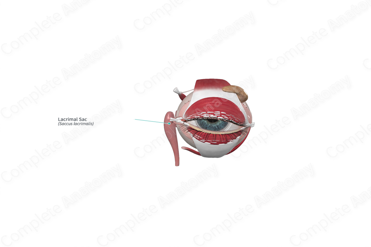
The lacrimal sac is the dilated superior beginning of the nasolacrimal duct located medially within the orbit; it receives the lacrimal canaliculi (Dorland, 2011).


Related parts of the anatomy
Structure and/or Key Feature(s)
The lacrimal sac is the bulbous, closed superior end of the nasolacrimal duct. The superior and inferior lacrimal canaliculi empty into it. The sac serves as a receptacle to collect tears before draining them down the nasolacrimal duct into the nasal cavity. A small slip of orbicularis oculi (the lacrimal portion) passes over the sac facilitating its emptying (Standring, 2016).
Anatomical Relations
The lacrimal collecting and drainage system, including the lacrimal sac is located at the medial angle of the eye. Here there is a bony fossa formed by the lacrimal bone (posteriorly) and the frontal process of the maxilla (anteriorly) in which it lies. The small lacrimal portion of orbicularis oculi passes across the ridges of the lacrimal fossa forming the anteromedial relation of the lacrimal sac. The sac continues inferiorly between the lacrimal and maxillary bones exiting the orbit as the nasolacrimal canal.
Function
The lacrimal sac is part of the apparatus for draining tears from the medial angle of the eye. The lacrimal canaliculi transport tears to the lacrimal sac, and then onto the nasolacrimal duct which directs the tears to the inferior meatus of the nasal cavity. If tear fluid accumulates faster than it can flow down the nasolacrimal duct, it collects here. Contraction of the lacrimal portion of orbicularis oculi (by blinking) can squeeze the sac, propelling the fluid at higher pressure and volume along the drainage system.
References
Dorland, W. (2011) Dorland's Illustrated Medical Dictionary. 32nd edn. Philadelphia, USA: Elsevier Saunders.
Standring, S. (2016) Gray's Anatomy: The Anatomical Basis of Clinical Practice. Gray's Anatomy Series 41 edn.: Elsevier Limited.
