Quick Facts
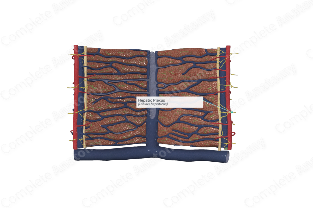
The hepatic plexus is an autonomic plexus accompanying the hepatic artery to the liver (Dorland, 2011).
Related parts of the anatomy
Structure/Morphology
The parenchyma of the liver receives both sympathetic and parasympathetic innervation via the hepatic plexus. The hepatic plexus receives preganglionic parasympathetic fibers originating from the anterior vagus trunk, with some contributions from the posterior vagal trunk, and postganglionic sympathetic fibers from the celiac and superior mesenteric plexuses (Standring, 2016).
Nerve fibers from the hepatic plexus enter the liver via the porta hepatis and accompany the portal triad into the parenchyma of the liver.
Anatomical Relations
The portal triad nerves lay on the surface of the vessels within the portal triad, winding around them and travelling alongside them through the hepatocytes in the hepatic lobule.
Function
Sympathetic fibers of the hepatic plexus innervate the blood vessels resulting in an increase in vascular resistance, a decrease in hepatic blood flow through the liver, and a rapid increase of glucose serum levels (Pawlina, 2016).
Parasympathetic fibers innervate the bile ducts (the larger ones with smooth muscle cells in their walls) and the blood vessels and promotes glucose uptake and utilization (Pawlina, 2016)
References
Dorland, W. (2011) Dorland's Illustrated Medical Dictionary. 32nd edn. Philadelphia, USA: Elsevier Saunders.
Pawlina, W. 2016. Histology: A text and atlas with correlated cell and molecular biology. 7th ed. Philadelphia: Wolters Kluwer.
Standring, S. (2016) Gray's Anatomy: The Anatomical Basis of Clinical Practice. Gray's Anatomy Series 41st edn.: Elsevier Limited.
