Quick Facts
The stratum basale, or basal layer of epidermis, is the deepest layer of the epidermis, beneath the stratum spinosum, composed of a single layer of cells, including stem cells, melanocytes, and Merkel cells. The stratum spinosum, or spinous layer, is the layer of epidermis between the stratum granulosum and stratum basale; it contains the prickle cells (Dorland, 2011).
Related parts of the anatomy
Structure/Morphology
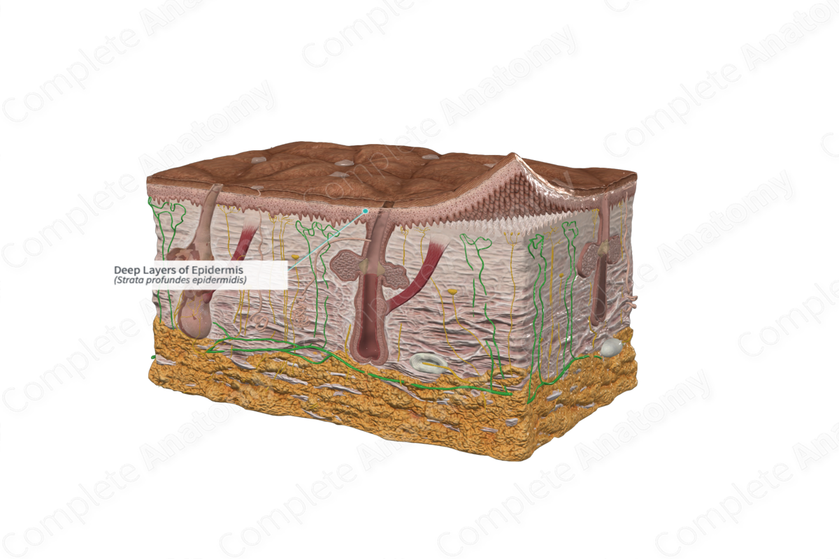
The deep layers of the epidermis are composed of the stratum spinosum and stratum basale. The stratum spinosum forms the thickest layer of the epidermis. It contains several layers of polyhedral keratinocytes, but cells at the most superficial surface of this layer are flattened. The processes of adjacent keratinocytes are connected by desmosomes, which adds to the strength of this layer. The stratum spinosum also contains Langerhans cells, or antigen-presenting dendritic cells. They play an important role in initiating immune response against foreign antigens (Arda, Goksugur and Tuzun, 2014).
The stratum basale is the deepest layer of the epidermis. It is composed of a single layer of cuboidal/columnar keratinocytes that are firmly attached to a basement membrane. Keratinocytes of the stratum basale are continuously proliferating from a stem cell population and are pushed into the overlying stratum spinosum.
A number of other cell populations are found in the stratum basale, including melanocytes and Merkel cells. Melanocytes are responsible for producing melanin, a pigment that accumulated in melanosomes released from the melanocytes (Arda, Goksugur and Tuzun, 2014). Keratinocytes take up these melanosomes, via phagocytosis, thus, releasing the melanin to the cytoplasm of the keratinocytes. Melanin protects the DNA of cells by absorbing ultraviolet, or UV, rays. Skin exposed to UV rays stimulates melanin production and accelerates its transfer to keratinocytes. Merkel cells are the tactile cells of the skin. They act as mechanoreceptors and play a role is sensory perception (Arda, Goksugur and Tuzun, 2014).
References
Arda, O., Goksugur, N. and Tuzun, Y. (2014) 'Basic histological structure and functions of facial skin', Clin Dermatol, 32(1), pp. 3-13.
Dorland, W. (2011) Dorland's Illustrated Medical Dictionary. 32nd edn. Philadelphia, USA: Elsevier Saunders.
