
Structure/Morphology
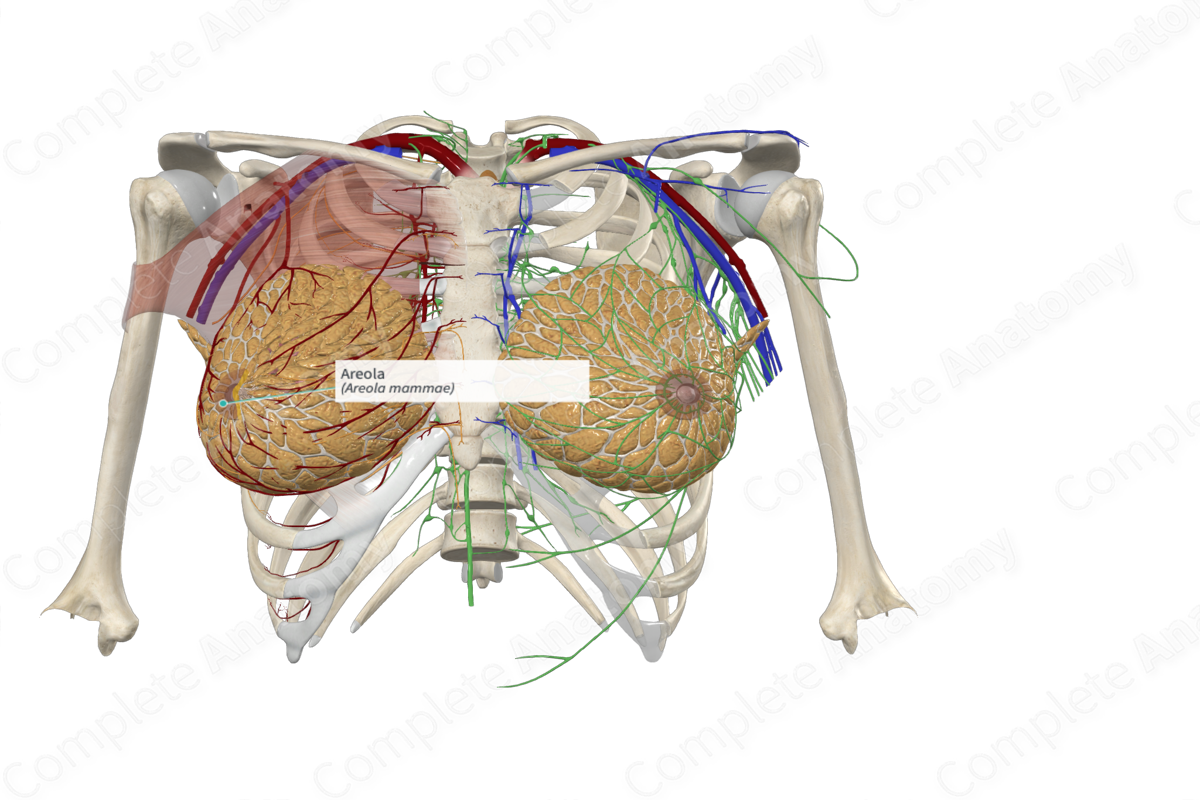
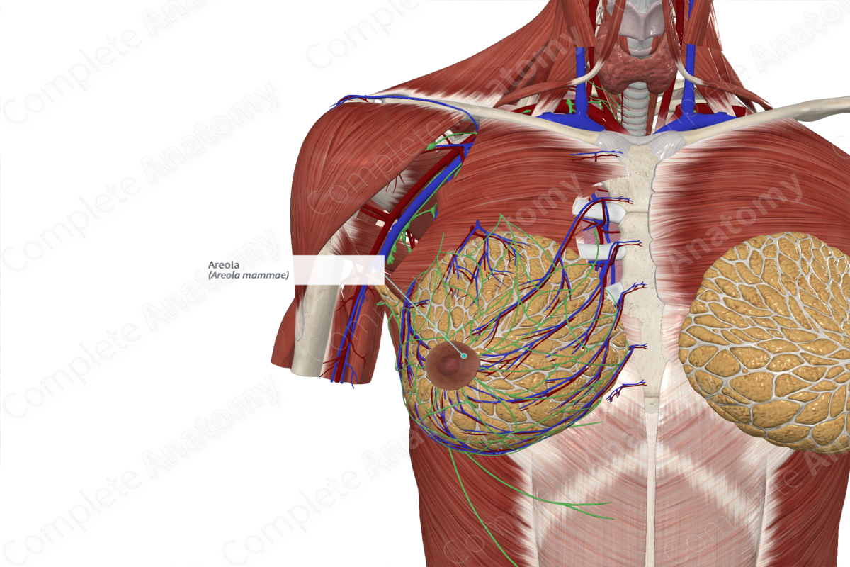
The areola is a highly pigmented, rounded tissue situated in the center of the breast. It is generally 3–6 cm in diameter. The nipple projects from the center of the areola (Zucca-Matthes, Urban and Vallejo, 2016).
The skin covering the areola is convoluted and shares and contains numerous sebaceous glands. The sebaceous glands of the areola are not associated with hair follicles. The openings of the sebaceous glands can be identified as projections on the surface of the areola, tubercles of Montgomery, and become enlarged during pregnancy.
The areola contains smooth muscle fibers that are arranged both radially (Meyerholz muscle) and circularly (Sappey muscle). This areolar muscle is continuous with the smooth muscle surrounding the lactiferous ducts in the nipple. Contraction of the muscle allows for secretion of the milk produced by the lactiferous glands (Zucca-Matthes, Urban and Vallejo, 2016).

Related parts of the anatomy
Key Features/Anatomical Relations
The areola is located at the level of the fourth intercostal space at the mid-clavicular line. However, this varies greatly, depending on the sex and age of the individual.
Function
The specialized arrangement of the sebaceous glands in the areola protects the skin of the breast from irritation and cracking during breast-feeding.
References
Zucca-Matthes, G., Urban, C. and Vallejo, A. (2016) 'Anatomy of the nipple and breast ducts', Gland Surgery, 5(1), pp. 32-36.
