Structure/Morphology
The anterior surface of the eyelids is covered by a modified layer of skin. It makes up about two thirds of the thickness of the eyelid, with the remainder composed of conjunctival mucosa. Between the skin and mucosa is a narrow “gray line” which marks the location of the ciliary bundle of the palpebral part of the orbicularis oculi muscle (the muscle of Riolan). This line allows for the separation of the eyelid across a relatively avascular plane into anterior and posterior portions (Standring, 2016).
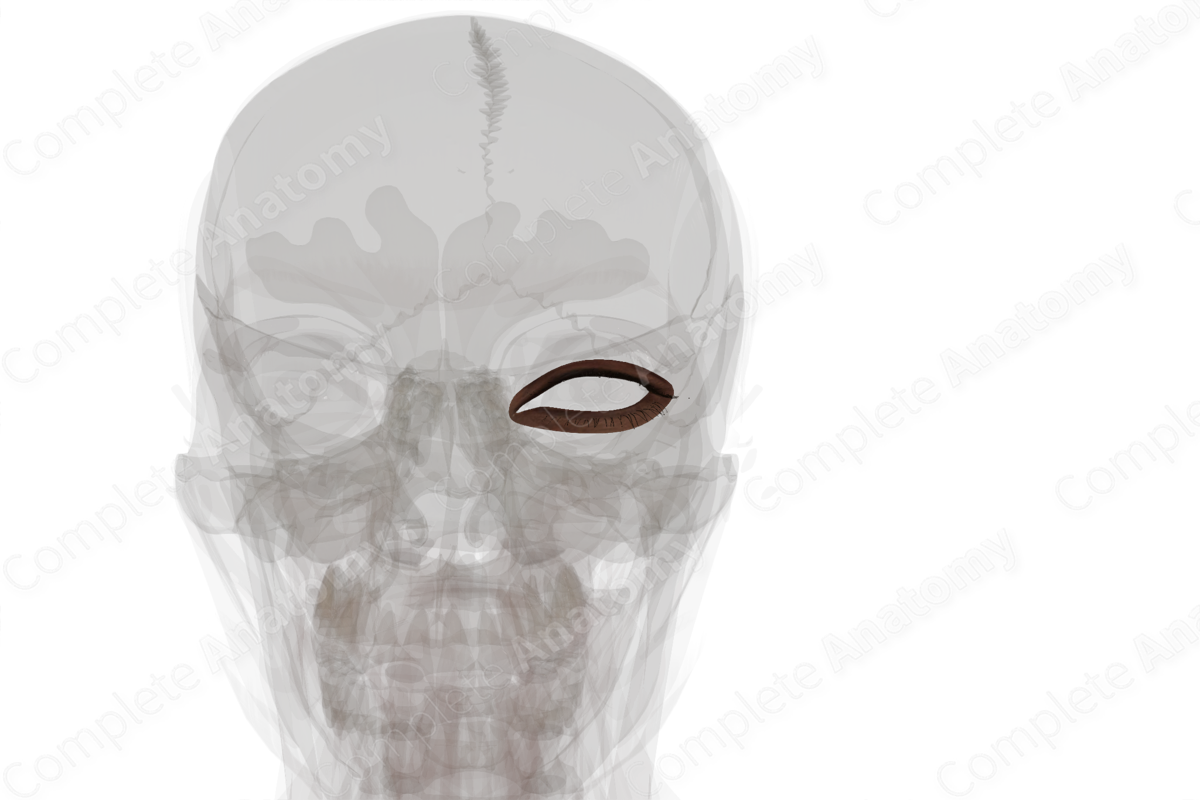
Sensory innervation of the upper eyelid arises from the ophthalmic division of the trigeminal nerve via the supraorbital branch of the frontal nerve. It may also receive contributions from the lacrimal and supratrochlear branches of the frontal nerve and from infratrochlear branches of the nasociliary nerve.
The lower eyelid receives sensory innervation from the infraorbital branch of the maxillary nerve, the second division of the trigeminal nerve.


Related parts of the anatomy
Key Features/Anatomical Relations
The skin of the eyelid contains numerous folds. The superior palpebral furrow lies opposite the margin of the superior tarsus and is deepened when the eyelids are open. The less prominent inferior palpebral furrow also lies opposite its corresponding tarsal plate, which deepens during downward gaze.
The skin around the medial angle of the eye varies between populations. In Caucasians, the medial angle of the eye is completely exposed, while in Asians, the medial canthus is covered by the epicanthus (a semilunar fold of skin).
Function
The eyelids protect the eye from mechanical trauma and serve as a shield from excessive light. Periodic blinking helps prevent corneal dryness (and ulceration) by maintaining a homogenous distribution of tear film over the cornea.
References
Standring, S. (2016) Gray's Anatomy: The Anatomical Basis of Clinical Practice. Gray's Anatomy Series 41 edn.: Elsevier Limited.
Learn more about this topic from other Elsevier products
Skin

Skin is a complex organ composed of two major layers—the epidermis and dermis—and a network of collagen, elastic fibers, blood vessels, and nerve endings.
