Quick Facts
Location: Perineum.
Arterial Supply: Internal and external pudendal arteries.
Venous Drainage: Internal and external pudendal veins.
Innervation: Somatic: pudendal nerve (S2—S4), ilioinguinal (L1) and genitofemoral nerve (L1-L2); Sympathetic: lumbar and sacral splanchnic nerves; Parasympathetic: pelvic splanchnic nerves.
Lymphatic Drainage: Paravaginal lymph nodes.
Structure/Morphology
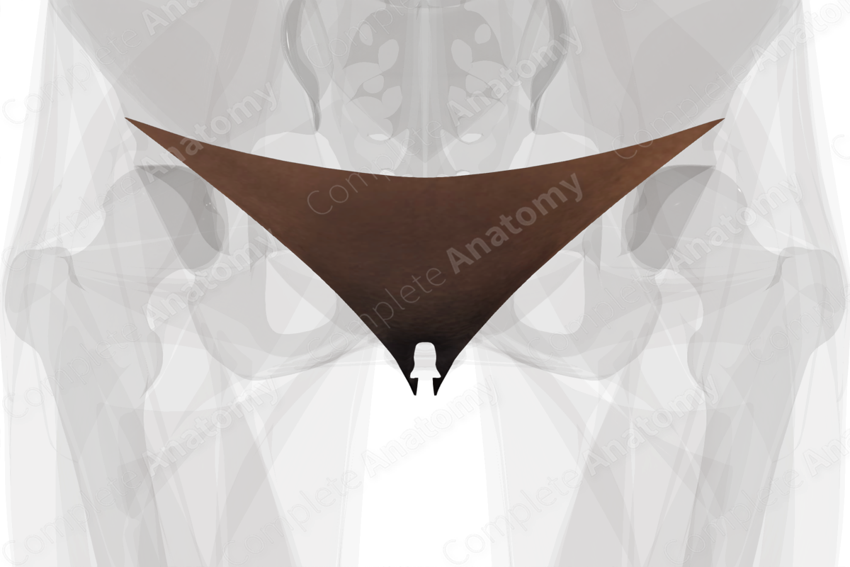
The vulva is the collective term that applies to the region containing the female external genitalia and includes the mons pubis, labia majora and minora, clitoris, vestibule as well as the bulb and the greater vestibular glands. In addition, it contains the urinary meatus.
Anatomical Relations
The vulva extends from the mons pubis anteriorly which overlies the pubic bone to the skin that overlies the perineal body posteriorly. The posterior location is approximately 2.5—3 cm anterior to the anus.
Function
The vulva consists of the external genitalia and is responsible for sexual arousal. Additionally, it protects the opening of the vaginal canal and urethra.
Arterial Supply
The vascular supply to the vulva comes from the internal pudendal artery, a branch from the internal iliac artery, which supplies the majority of the inferior portion of the vulva. The external pudendal artery, a branch of the femoral artery, supplies the majority of the superior portion of the vulva.
Venous Drainage
Venous blood from the skin of the vulva predominately drains into the external pudendal vein to the long saphenous vein. Venous blood from the clitoris drains into the deep dorsal veins of the clitoris to the internal pudendal veins and the internal iliac veins and via superficial dorsal veins to the external pudendal and long saphenous veins.
Innervation
The vulva receives somatic innervation from the pudendal nerve (S2—S4) via the inferior rectal branch, the perineal branch and the dorsal nerve of the clitoris. Sensory innervation to the anterior third of the labia majora is from the ilioinguinal nerve (L1) while the remainder is supplied by the pudendal nerve via the posterior labial branch of the perineal nerve (S3). Additional innervation is supplied by the perineal branch of the posterior cutaneous nerve of the thigh (S2), and the genitofemoral nerve.
The organs of the vulva also receive autonomic innervation through the splanchnic nerves, that synapse in the superior and inferior hypogastric/mesenteric plexuses. Sympathetic nerve supply is supplied through the lumbar and sacral splanchnic nerve (L1—L2).
Parasympathetic nerve innovation to the genitals is supplied through the pelvic splanchnic nerves (S2—S4). The splanchnic nerves (Standring, 2016).
Lymphatic Drainage
The paravaginal lymph nodes are located around the vagina. The lowest nodes of this group drain lymph from vulva and perineal skin, which passes to the superficial inguinal lymph nodes (Földi et al., 2012). Additionally, lymph from the lower labia majora is returned to the rectal lymph plexus, while lymph from the clitoris drains direct to the deep inguinal lymph nodes (Standring, 2016).
List of Clinical Correlates
- Sexually Transmitted Diseases (STDs)
- Vulvar cancer
- Vulvar pain syndrome
- Vulvodynia
- Childbirth
References
Földi, M., Földi, E., Strößenreuther, R. and Kubik, S. (2012) Földi's Textbook of Lymphology: for Physicians and Lymphedema Therapists. Elsevier Health Sciences.
Standring, S. (2016) Gray's Anatomy: The Anatomical Basis of Clinical Practice. Gray's Anatomy Series: Elsevier Limited.


