Quick Facts
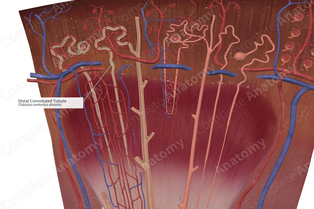
The distal convoluted tubule is a distal, convoluted part of the renal tubule, extending from the distal straight tubule to the connecting tubule (Dorland, 2011).
Structure and/or Key Features
The distal convoluted tubule is considered the final segment of the renal tubule. Measuring approximately 30–50 µm in diameter, the distal convoluted tubule is characterized by a lower height cuboidal epithelium, no brush border of microvilli, and a generally wider lumen (Kerr, 2007).
The point where the distal convoluted tubule continues on from the distal straight tubule, or thick ascending limb, is demarcated by a region of narrowed, clustered cells, known as the macula densa.
Anatomical Relations
Along the renal tubule, the distal convoluted tubule succeeds the thick ascending limb and macula densa.
Function
The distal convoluted tubule is responsible for the selective reabsorption of ions, such as Na+ and Ca2+, and water from the tubular fluid. Na+ is transported through Na+/K+ ATPase pumps. Na+ reabsorption along the distal convoluted tubule is mediated by the hormone aldosterone, which increases the number of Na+/K+ ATPase pumps. K+, on the other hand, is secreted along the distal convoluted tubule (Pocock et al, 2013).
The distal convoluted tubule regulates pH by absorbing and secreting both bicarbonate and protons (H+) along its length. It is also responsible for the secretion of acids, ammonia, drugs, and toxins (Martini et al, 2017; Pocock et al, 2013).
List of Clinical Correlates
—Gitelman syndrome
—Gordon syndrome
References
Dorland, W. (2011) Dorland's Illustrated Medical Dictionary. 32nd edn. Philadelphia, USA: Elsevier Saunders.
Kerr, J. B. (2007) Atlas of Functional HistologyElsevier Science Health Science Division.
Martini, F. H., Nath, J. L. & Bartholomew, E. F. (2017) Fundamentals of Anatomy & PhysiologyPearson Education.
Pocock, G., Richards, C. D. & Richards, D. A. (2013) Human Physiology, 4 edition. OUP Oxford.
