Quick Facts
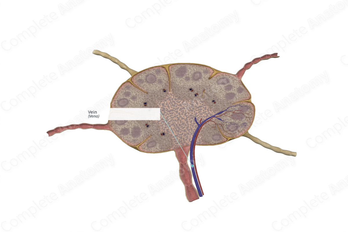
A vein is a vessel through which blood passes from various organs or parts back to the heart; all veins except the pulmonary veins carry blood low in oxygen. Like arteries, veins have three coats, an inner, middle, and outer, but the coats are not so thick, and they collapse when the vessel is cut. Many veins have valves formed of reduplications of their lining membrane, which prevent the backward flow of blood away from the heart (Dorland, 2011).
Structure/Morphology
Blood from the capillary beds of the cortex drain into the medullary venules within the medullary cords. These venules unify with other venules from other lobules of the lymph node, forming a vein which exists the lymph node at the hilum, along with the efferent lymph vessel and accompanying artery. Each lobule of the lymph node is drained by a vein, however, for descriptive purposes this is not shown in the 3D model.
Lymphocytes are capable of migrating from the venules into the paracortex, or deep cortex, through high endothelial venules. High endothelial venules are so-called due to the specialized characteristics of the endothelial cells in that part of the venules. Instead of a squamous endothelial cell, as seen in regular arteries and veins, and lymphatic vessels, the endothelial cells are cuboidal. They are capable of binding to intravascular lymphocytes and facilitate the passage of the cells from the blood into the reticular tissue of the paracortex. In fact, most lymphocytes enter the lymph nodes through high endothelial venules (Willard-Mack, 2006; Miyasaka and Tanaka, 2004).
As the venule continues into the medullary region of the lymph node, it loses its high endothelium, which has reverted to the squamous endothelium.
References
Dorland, W. (2011) Dorland's Illustrated Medical Dictionary. 32nd edn. Philadelphia, USA: Elsevier Saunders.
Miyasaka, M. and Tanaka, T. (2004) 'Lymphocyte trafficking across high endothelial venules: dogmas and enigmas', Nat Rev Immunol, 4(5), pp. 360-70.
Willard-Mack, C. L. (2006) 'Normal structure, function, and histology of lymph nodes', Toxicologic Pathology, 5(34), pp. 409-424.
