Quick Facts
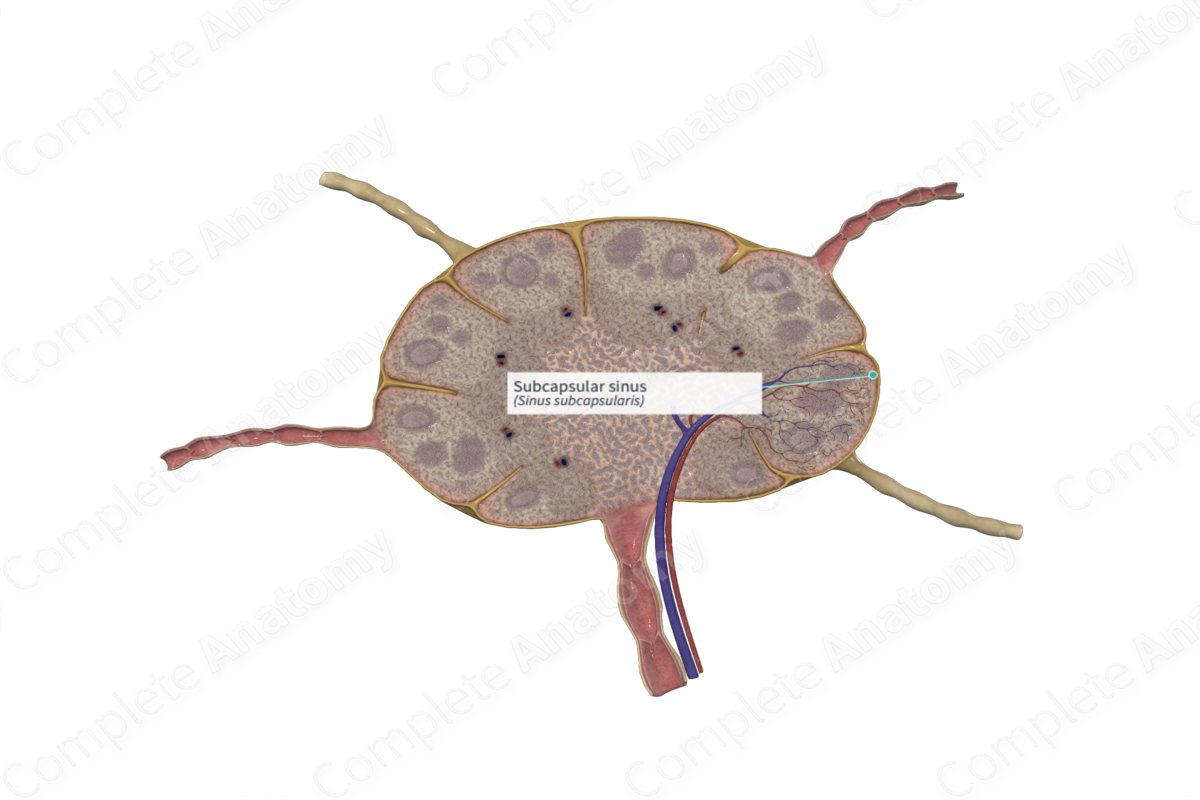
The subcapsular sinuses are the bowl-shaped lymph sinuses separating the capsule from the cortical parenchyma of a lymph node, and from which lymph flows into the cortical sinuses (Dorland, 2011).
Structure/Morphology
The subcapsular sinus is the area where afferent lymph vessels empty their lymph into. Unlike vascular sinuses, lymphatic sinuses are not open spaces. They are occupied by reticular fibers surrounded by reticular cell processes and macrophages. This forms a meshwork within the sinus, thus slowing down the flow of lymph through the sinus which enhances filtration.
The subcapsular sinus is one of three sinuses in the lymph node, the others include the trabecular (or transverse) and medullary sinuses.
Anatomical Relations
The subcapsular sinus separates the lymphocyte-rich cortex of the lymph node from its capsule. An afferent lymph vessel enters the lymph node through the capsule and supplies a constant supply of lymph to the subcapsular sinus. Lymph passes from the subcapsular sinus to the trabecular sinus and then the medullary sinus, where it collects at an efferent lymph vessel on the hilar side of the lymph node.
Function
The subcapsular sinus receives lymph from the afferent lymph vessels. The lymph is filtered by the reticular meshwork in the sinus, where the antigenic material becomes trapped and phagocytosed by the macrophages (Willard-Mack, 2006).
References
Dorland, W. (2011) Dorland's Illustrated Medical Dictionary. 32nd edn. Philadelphia, USA: Elsevier Saunders.
Willard-Mack, C. L. (2006) 'Normal structure, function, and histology of lymph nodes', Toxicologic Pathology, 5(34), pp. 409-424.
