Quick Facts
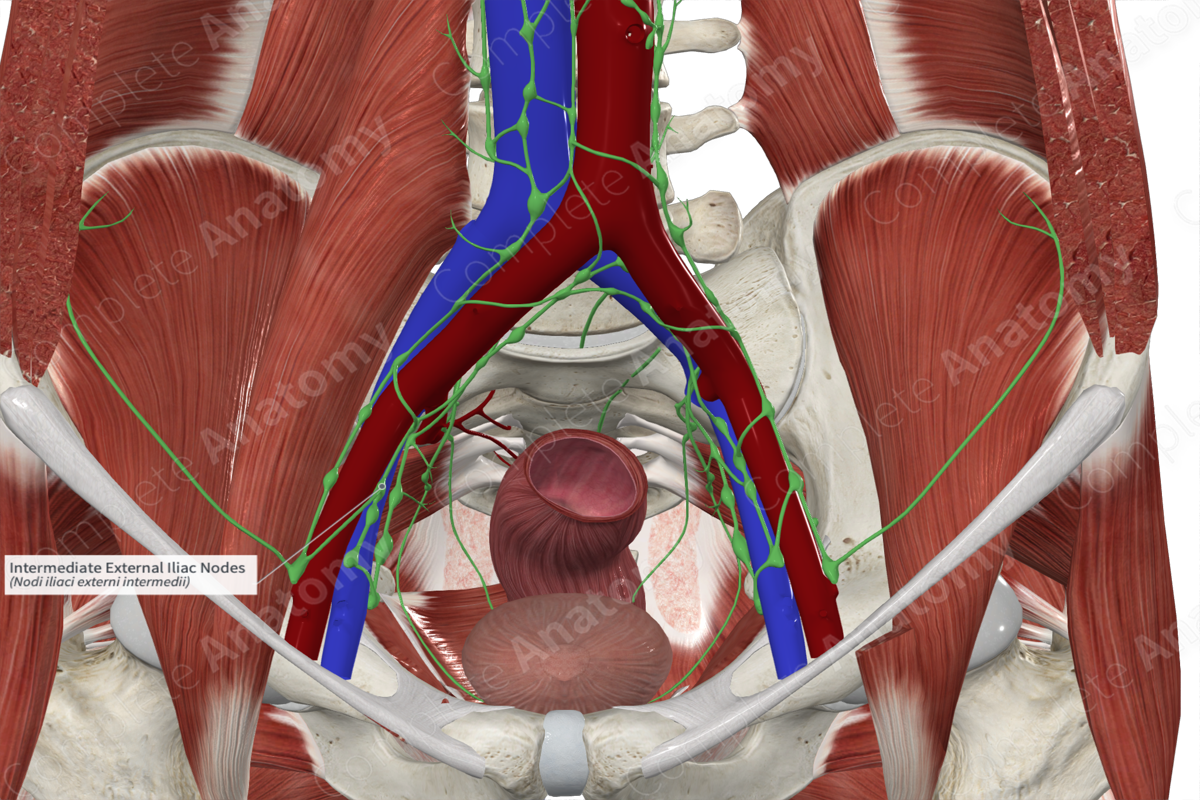
Location: Between the external iliac artery and vein.
Drainage: Deep layers of the abdominal wall, viscera of pelvis, perineum, and lower limb via the inguinal lymph nodes.
Direction of Flow: Common iliac lymph nodes > Lateral aortic lymph nodes (left) and lateral caval lymph nodes (right) > left and right lumbar lymph trunk > cisterna chyli > thoracic duct.


Description:
Description: (Location & Drainage)
The intermediate external iliac lymph nodes are located between the external iliac artery and vein. This group is often absent in some individuals (20%) (Földi et al., 2012).
This group usually consists of two or three nodes, with the superior node of this group most likely to be present (60%) (Földi et al., 2012). The superior node is located between the external and internal iliac arteries and is also referred to as the interiliac node along with the uppermost node of the medial external iliac lymph nodes. The interiliac nodes are important as they act as a confluence to the external and internal iliac lymphatic pathways.
The most inferior node of the intermediate external iliac lymph nodes is called the intermediate lacunar node and is often absent.
The intermediate external iliac lymph nodes receive lymph from the viscera of the pelvis including the fundus of the bladder, urethra, cervix, superior part of the vagina and prostate. The intermediate lacunar lymph node of this group receives lymph from the deep layers of the abdominal wall, inferior to the umbilicus, the perineum, and lower limb via the inguinal lymph nodes.
The intermediate external iliac lymph nodes send efferent lymph vessels to the intermediate and medial common iliac lymph nodes, which send lymph to the lateral aortic and caval lymph nodes of the left and right side, respectively.
References
Földi, M., Földi, E., Strößenreuther, R. and Kubik, S. (2012) Földi's Textbook of Lymphology: for Physicians and Lymphedema Therapists. Elsevier Health Sciences.
Description:
Description: (Location & Drainage)
The intermediate external iliac lymph nodes are located between the external iliac artery and vein. This group is often absent in some individuals (20%) (Földi et al., 2012).
This group usually consists of two or three nodes, with the superior node of this group most likely to be present (60%) (Földi et al., 2012). The superior node is located between the external and internal iliac arteries and is also referred to as the interiliac node along with the uppermost node of the medial external iliac lymph nodes. The interiliac nodes are important as they act as a confluence to the external and internal iliac lymphatic pathways.
The most inferior node of the intermediate external iliac lymph nodes is called the intermediate lacunar node and is often absent.
The intermediate external iliac lymph nodes receive lymph from the viscera of the pelvis including the fundus of the bladder, urethra, cervix, superior part of the vagina and prostate. The intermediate lacunar lymph node of this group receives lymph from the deep layers of the abdominal wall, inferior to the umbilicus, the perineum, and lower limb via the inguinal lymph nodes.
The intermediate external iliac lymph nodes send efferent lymph vessels to the intermediate and medial common iliac lymph nodes, which send lymph to the lateral aortic and caval lymph nodes of the left and right side, respectively.
Learn more about this topic from other Elsevier products
External Iliac Lymph Nodes

The external iliac lymph node chain is typically comprised of 9-10 lymph nodes forming 3 distinct chains of approximately 3 nodes each, called the lateral, middle/intermediate, and medial chains.
