Quick Facts
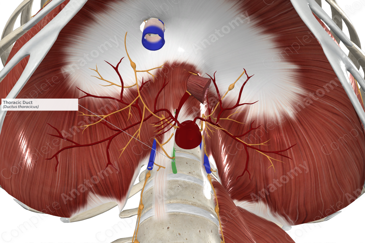
Location: Sits on the vertebral bodies.
Drainage: Structures below the diaphragm and from the left side above the diaphragm.
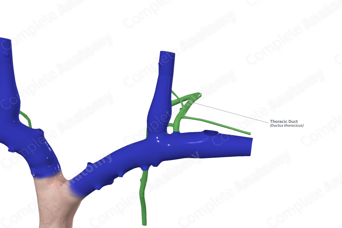
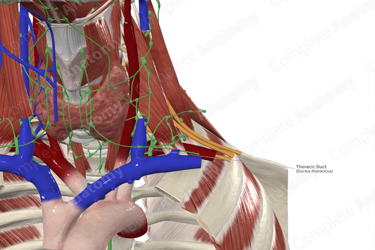
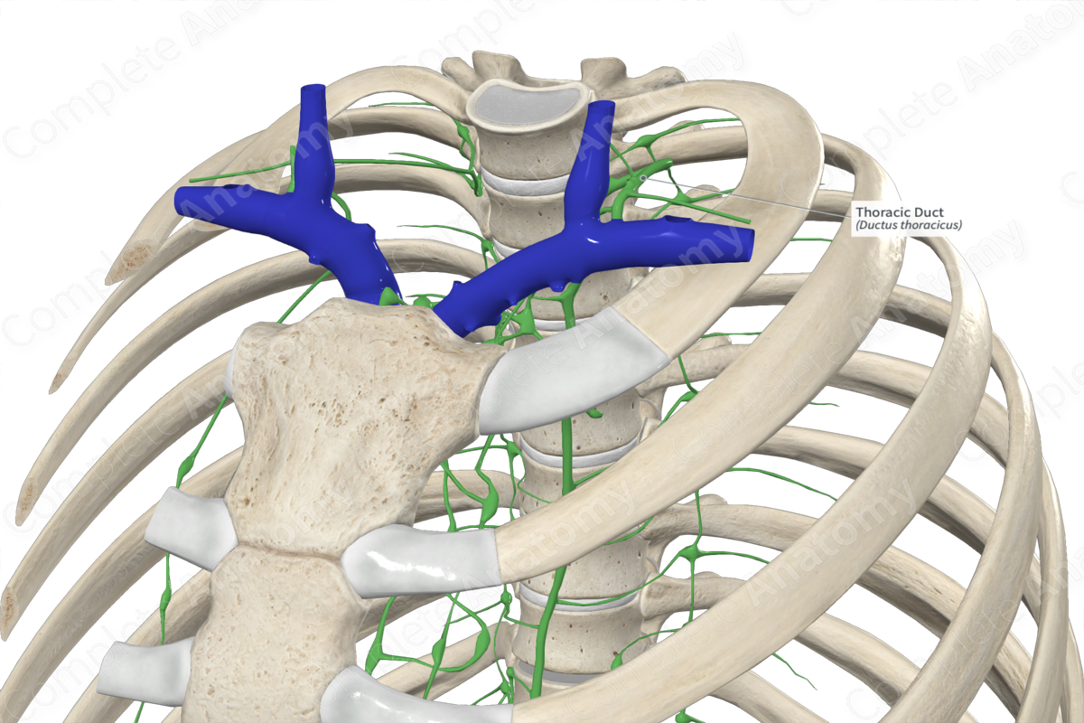
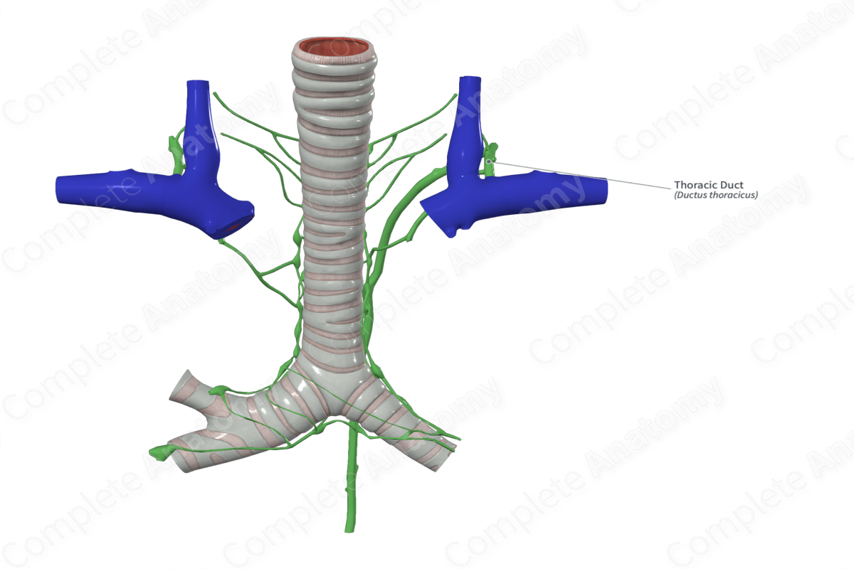
Direction of Flow: Drains into the venous system at the junction of the left subclavian and internal jugular veins.
Related parts of the anatomy
Description: (Location & Drainage)
The thoracic duct is the largest lymph vessel in the body and forms the superior extension of the cisterna chyli. The duct originates in the lumbar region, at about the level of the second lumbar vertebra, although this varies significantly depending on body position and how distended the cisterna chyli is.
As the thoracic duct ascends briefly in the lumbar region, it receives the descending efferent vessels of the intercostal lymph nodes (along with the intercalated prevertebral nodes). It ascends into the thorax, through the aortic hiatus, and continues on the left side of the esophagus. As it reaches the superior aperture of the thorax, it arches over the subclavian artery to empty into the venous system at the junction of the left subclavian and internal jugular veins. The thoracic duct has numerous paired semilunar valves, usually 1–8, but can contain as many as twenty. This gives the duct a beaded appearance.
List of Clinical Correlates
—Thoracic duct laceration
—Thoracic duct rupture
Learn more about this topic from other Elsevier products
Thoracic Duct

The thoracic duct drains lymph from most of the body and the left side of the head into the venous system at the junction of the left internal jugular and subclavian veins.

