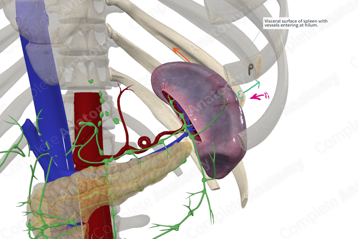
Quick Facts
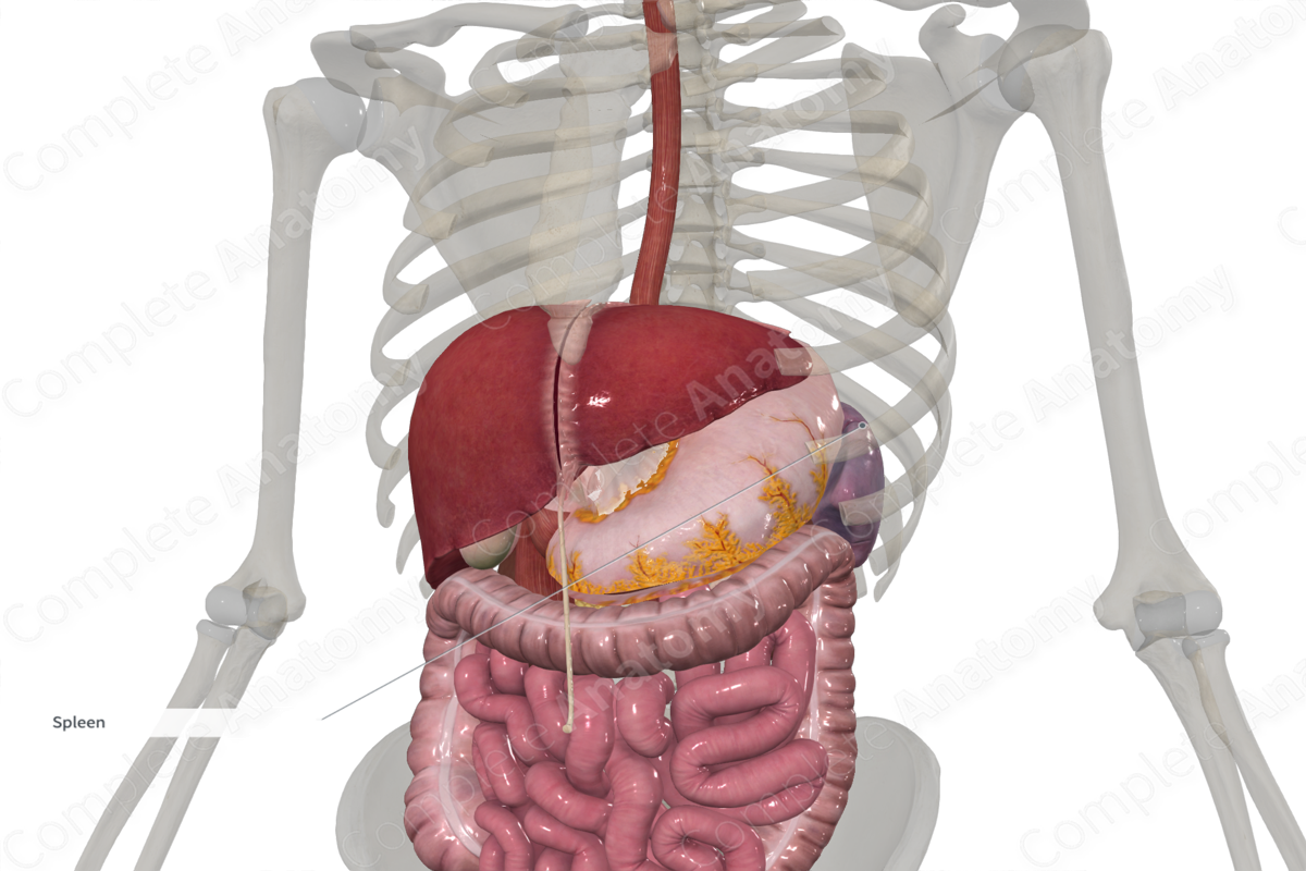
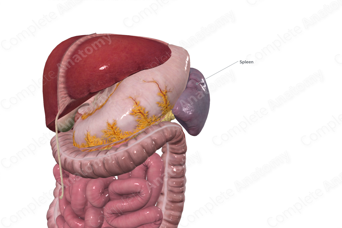
Location: Left epigastric region of abdomen.
Arterial Supply: Splenic artery.
Venous Drainage: Splenic vein.
Innervation: Sympathetic: postganglionic fibers from the celiac ganglion; Parasympathetic: preganglionic fibers from vagal trunks.
Lymphatic Drainage: Splenic nodes > superior pancreatic nodes > celiac nodes > intestinal trunk > cisterna chyli > thoracic duct.
Related parts of the anatomy
Structure
The spleen is classified as a secondary lymph organ and is the largest lymph organ in the body. It has a convex, smooth lateral or external surface. A sharp border, notched anteriorly, separates this surface from the concave medial or internal surface. The later forms the hilum for the entry of neural, vascular, and lymphatics to the organ. The hilum is surrounded by impressions left by other abdominal organs to which it is closely related.
Anatomical Relations
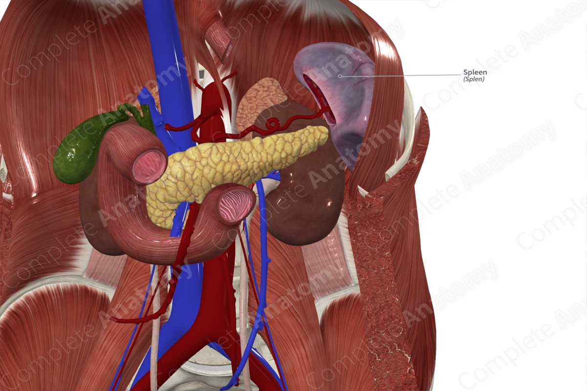
The spleen is located in the left epigastric region of the abdomen. The lateral surface of the spleen lies just inferior to the far-left side of the diaphragm, just medial to the left costodiaphragmatic recess. The impressions on its medial surface are formed superiorly by the fundus of the stomach, inferiorly by the left colic flexure (hence its alternate name “splenic flexure”), and posteriorly by the superior pole of the kidney and its surrounding perirenal fat pad. The very distal end of the tail of the pancreas is also very closely related to the medial surface of the spleen, at the hilum.
A mnemonic that helps in recalling the metrics of the spleen is the 1x3x5x7x9x11 rule. This rule describes the spleen as being 1" thick, 3" wide by 5" long, weighs around 7 oz (200 grams), and lies between the 9th and 11th ribs.
Function
The functions of the spleen are many and still incompletely understood. It is primarily an organ of the immune system and as such plays an important role in immunological defense. It is also known to be involved in metabolism and maintenance of some components of the blood.
Arterial Supply
The splenic artery, one of the three primary branches of the celiac trunk, provides the arterial supply to the spleen.
Venous Drainage
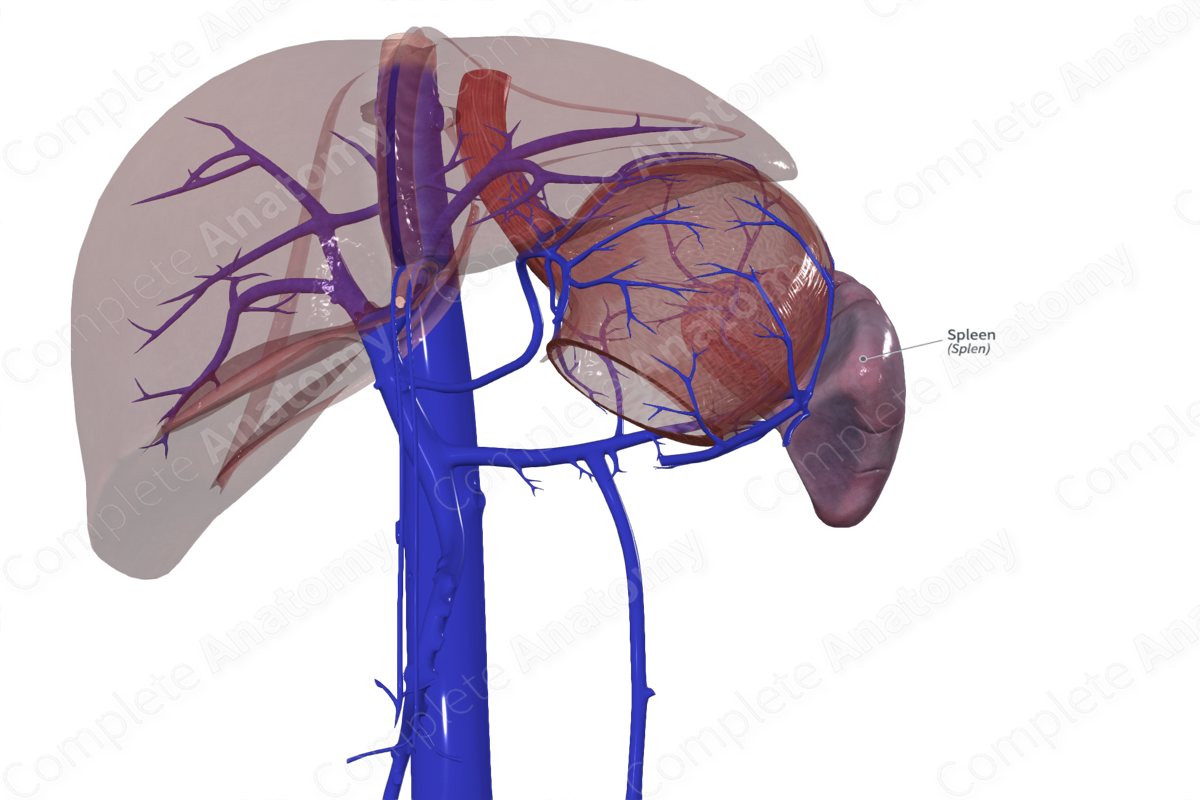
The splenic vein drains the spleen. It typically joins the inferior mesenteric vein just before it merges with the superior mesenteric vein to form the portal vein.
Innervation
The spleen is innervated by both divisions of the autonomic nervous system that forms a plexus around its vasculature. This plexus includes postganglionic sympathetic fibers originating in the celiac ganglion and preganglionic parasympathetics from the vagal trunks.
Lymphatic Drainage
Lymphatics travel with blood vessels collecting into a plexus that drains into lymph nodes of the splenic hilum and pancreatic tail. From here, lymph drains to superior pancreatic nodes, as well as inferior pancreatic and left gastroomental lymph nodes, eventually draining into celiac nodes and the cisterna chyli (Földi et al., 2012).
List of Clinical Correlates
—Hemopoiesis
—Splenectomy
References
Földi, M., Földi, E., Strößenreuther, R. and Kubik, S. (2012) Földi's Textbook of Lymphology: for Physicians and Lymphedema Therapists. Elsevier Health Sciences.
Learn more about this topic from other Elsevier products
Spleen histology: Video, Causes, & Meaning

Spleen histology: Symptoms, Causes, Videos & Quizzes | Learn Fast for Better Retention!

