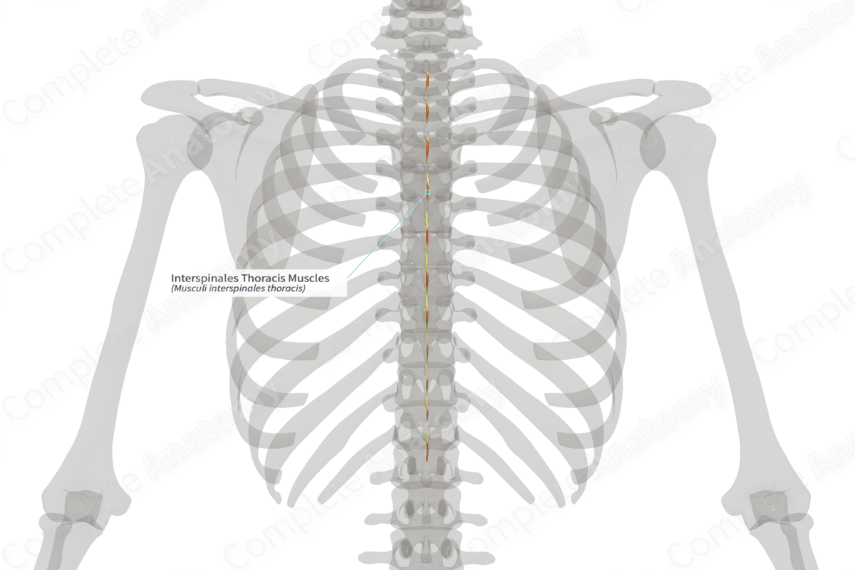
Interspinales Thoracis Muscles (Right) Quick Facts
Origin: Superior aspects of spinous processes of T2-T12 vertebrae.
Insertion: Inferior aspects of spinous processes of T1-T11 vertebrae.
Action: Extends trunk.
Innervation: Posterior rami of thoracic nerves.
Arterial Supply: Dorsal branches of posterior intercostal arteries.


Related parts of the anatomy
Interspinales Thoracis Muscles (Right) Origin
The interspinales thoracis muscles originate from the superior aspects of the spinous processes of the second to twelfth thoracic vertebrae. There can be variations between individuals regarding the origin sites for the interspinales thoracis muscles (Tubbs, Shoja and Loukas, 2016).
Interspinales Thoracis Muscles (Right) Insertion
The fibers of the interspinales thoracis muscles travel superiorly to the thoracic vertebrae that are located one vertebral segment superior to their origin sites. They insert onto the inferior aspects of the spinous processes of the first to eleventh thoracic vertebrae. There can be variations between individuals regarding the insertion sites for the interspinales thoracis muscles (Tubbs, Shoja and Loukas, 2016).
Interspinales Thoracis Muscles (Right) Key Features & Anatomical Relations
Overall, the interspinales (interspinal) muscles are one of three groups of muscles found in the deep layer of the intrinsic muscles of the back. This group of muscles is collectively known as the “interspinales muscles” because they originate from and insert onto the spinous processes of adjacent vertebrae.
They are well developed in the cervical and lumbar regions and less so in the thoracic region and are made up of many short, thin skeletal muscles. For classification purposes, the interspinales muscles are divided into three parts:
- interspinales colli, which is the superior portion;
- interspinales thoracis, which is the middle portion;
- interspinales lumborum, which is the inferior portion.
The interspinales muscles are located:
- medial to the erector spinae, transversospinal and spinotransversales muscles;
- lateral to the interspinous ligaments.
Interspinales Thoracis Muscles (Right) Actions
The interspinales thoracis muscles extend the trunk (Moore, Dalley and Agur, 2009). They may also be involved in proprioception (Standring, 2016).
Interspinales Thoracis Muscles (Right) References
Moore, K. L., Dalley, A. F. and Agur, A. M. R. (2009) Clinically Oriented Anatomy. Lippincott Williams & Wilkins.
Standring, S. (2016) Gray's Anatomy: The Anatomical Basis of Clinical Practice. Gray's Anatomy Series 41st edn.: Elsevier Limited.
Tubbs, R. S., Shoja, M. M. and Loukas, M. (2016) Bergman's Comprehensive Encyclopedia of Human Anatomic Variation. Wiley.
