Quick Facts
Origin: Area of maxilla superior to lateral incisor and canine teeth.
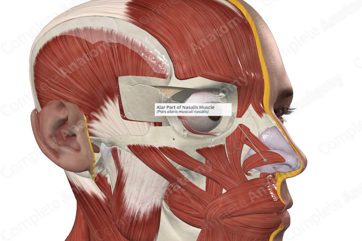
Insertion: Skin overlying lateral crus of major alar cartilage.
Action: Dilates nostrils.
Innervation: Buccal branches of facial nerve (CN VII).
Arterial Supply: Superior labial and infraorbital arteries.


Related parts of the anatomy
Origin
The nasalis muscle is divided into transverse and alar parts. The alar part of the nasalis muscle also arises from the maxilla, superior to the lateral incisor and canine teeth.
Insertion
The alar part of the nasalis muscle attaches to the skin overlying the lateral crus of the major alar cartilage and the posterior part of the mobile septum.
Actions
The alar part of the nasalis muscle dilates the nostrils by drawing the nasal septum downwards (Standring, 2016).
List of Clinical Correlates
- Bell’s palsy
References
Standring, S. (2016) Gray's Anatomy: The Anatomical Basis of Clinical Practice. Gray's Anatomy Series 41st edn.: Elsevier Limited.
Learn more about this topic from other Elsevier products
Alar Plate

The sulcus limitans is a groove that divides the more dorsal alar plate from the ventral basal plate.