Quick Facts
Origin: Common tendinous ring.
Insertion: Lateral surface of sclera.
Action: Abducts eyeball.
Innervation: Abducens nerve (CN VI).
Arterial Supply: Ophthalmic and lacrimal artery.
Origin
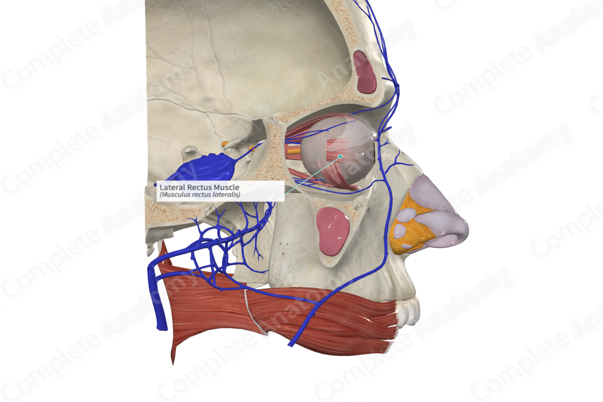
The lateral rectus muscle arises from the upper and lower lateral parts of the common tendinous ring. Additionally, some muscle fibers arise from the greater wing of the sphenoid.
Insertion
The lateral rectus muscle passes anteriorly in a horizontal plane along the lateral wall of the orbit. It inserts onto the lateral surface of the sclera just posterior to the corneoscleral junction.
Actions
The lateral rectus muscle works antagonistically with the medial rectus muscle to rotate the eyeball around the vertical axis. For lateral rectus, this means lateral rotation (abduction) exclusively. Working bilaterally this diverges the eyeballs when attention is drawn to objects that are distant.
List of Clinical Correlates
- Abducens nerve palsy
- Oculomotor nerve palsy
Learn more about this topic from other Elsevier products
Lateral Rectus Muscle

The lacrimal nerve is the smallest branch and courses along the upper border of the lateral rectus muscle to the lacrimal gland and then down to the conjunctiva and skin of the upper eyelid.


