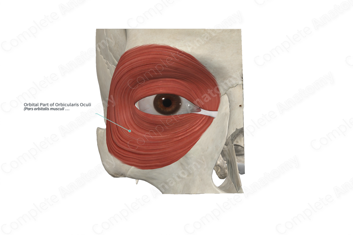
Quick Facts
Origin: Nasal part of frontal bone, frontal process of maxilla, and medial palpebral ligament.
Insertion: Adjacent muscles.
Action: Closes eyelids.
Innervation: Temporal and zygomatic branches of facial nerve (CN VII).
Arterial Supply: Facial, superficial temporal, maxillary, and ophthalmic arteries.
Related parts of the anatomy
Origin
The orbicularis oculi muscle is a broad, flat muscle that encircles the orbit, thus forming a sphincter around each of the eye sockets. It may be divided into three portions:
- orbital part;
- palpebral part;
- lacrimal part.
The orbital part of the orbicularis oculi muscle originates from the nasal part of the frontal bone, the frontal process of maxilla, and the medial palpebral ligament.
Insertion
The orbital part of the orbicularis oculi muscle encircles the orbit and its fibers blends with adjacent muscles.
Actions
The orbital part of the orbicularis oculi muscle closes the eyelids (Standring, 2016).
List of Clinical Correlates
- Bell’s palsy
- Lagophthalmos
References
Standring, S. (2016) Gray's Anatomy: The Anatomical Basis of Clinical Practice. Gray's Anatomy Series 41st edn.: Elsevier Limited.
Learn more about this topic from other Elsevier products


