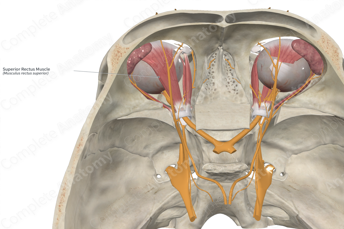
Quick Facts
Origin: Common tendinous ring.
Insertion: Upper surface of sclera.
Action: Elevates, adducts, and medially rotates eyeball.
Innervation: Superior branch of oculomotor nerve (CN III).
Arterial Supply: Ophthalmic and supraorbital artery.
Origin
The superior rectus muscle arises from the upper part of the common tendinous ring, superolateral to the optic canal. Additionally, some fibers of the superior rectus muscle arise from the dural sheath surrounding the optic nerve.
Insertion
From the common tendinous ring, the superior rectus muscle runs anterolaterally to inset obliquely on the upper part of the sclera.
Key Features & Anatomical Relations
Superior to the superior rectus muscle is the muscle belly of levator palpebrae superioris and inferior is the periorbital fat and the superior surface of the ocular globe.
Actions
The superior rectus muscle is primarily involved in elevating the eyeball around the transverse (horizontal) axis, i.e., moves the pupil superiorly. It does so primarily with the eye abducted. It is also involved in medial rotation (adduction) around the vertical axis and medial rotation (intorsion) around the anteroposterior axis, especially when the eye is adducted.
Additionally, a check ligament extends from superior rectus muscle to levator palpebrae superioris muscle, which ensures elevation of the superior eyelid when the pupil is elevated.
List of Clinical Correlates
- Diplopia
Learn more about this topic from other Elsevier products


