Quick Facts
Origin: Head and lateral surface of fibula, adjacent intermuscular septa.
Insertion: Plantar surface of medial cuneiform bone and base of first metatarsal bone.
Action: Everts foot at subtalar and transverse tarsal joints; assists in plantarflexion of foot at ankle joint.
Innervation: Superficial fibular nerve (L5-S1).
Arterial Supply: Anterior tibial and fibular arteries.


Related parts of the anatomy
Origin
The fibularis longus muscle originates from the:
- lateral aspect of the head of fibula;
- proximal half of lateral surface of fibula;
- adjacent intermuscular septum and crural fascia.
Insertion
The fibers of the fibularis longus muscle travel inferiorly to the foot and insert, via a long tendon, onto the:
- plantar surface of medial cuneiform bone;
- plantar aspect of the base of first metatarsal bone.
Key Features & Anatomical Relations
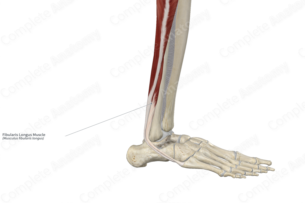
The fibularis longus (peroneus longus) muscle is found in the lateral compartment of the leg. It is a long, narrow, bipennate type of skeletal muscle. In the distal one third of the leg, the muscle belly gives rise to a tendon that travels inferiorly towards the malleolar groove of the fibula. The tendon hooks around the groove, changing its line of pull to a more anteroinferior direction. This tendon then travels deep to the superior and inferior fibular retinacula, posterior to the adjacent tendon of fibularis brevis, where it passes through the common tendinous sheath of fibularis muscles.
Along the lateral aspect of the foot, the tendon continues to travel anteroinferiorly, passing along the groove for tendon of fibularis longus muscle on the calcaneus, which is located inferior to the fibular trochlea of calcaneus. The tendon then travels towards the groove for tendon of fibularis longus muscle on the cuboid bone, which it will hook around and then change its line of pull to a more anteromedial direction. Within the plantar part of the foot, the tendon travels anteromedially to its insertion site.
The fibularis longus muscle is located:
- anterior to the soleus and flexor hallucis longus muscles;
- posterior to the fibularis brevis and extensor digitorum longus muscles
Actions & Testing
The fibularis longus muscle is involved in multiple actions:
- everts the foot at the subtalar and transverse tarsal joints;
- assists in plantarflexion of the foot at the ankle joint;
- helps stabilize the longitudinal and transverse arches of the foot.
The fibularis longus muscle can be tested by everting at the subtalar and transverse tarsal joints against resistance, during which its tendon can be seen and palpated (Standring, 2016).
List of Clinical Correlates
- Pes cavus
References
Standring, S. (2016) Gray's Anatomy: The Anatomical Basis of Clinical Practice. Gray's Anatomy Series 41st edn.: Elsevier Limited.
