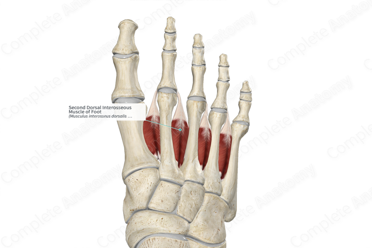
Second Dorsal Interosseous Muscle of Foot
Musculus interosseus dorsalis pedis secundus
Read moreQuick Facts
Origin: Lateral aspect of body of second metatarsal bone and medial aspect of body of third metatarsal bone.
Insertion: Lateral aspect of base of proximal phalanx of second toe and extensor expansion of second toe.
Action: Laterally abducts second toe at its metatarsophalangeal joint; simultaneously flexes metatarsophalangeal joint and extends interphalangeal joints of second toe.
Innervation: Deep branch of lateral plantar nerve (S2-S3).
Arterial Supply: Arcuate, dorsal and plantar metatarsal, and dorsal digital arteries of foot.


Origin
The second dorsal interosseous muscle of foot consists of two heads:
- the medial head, which originates from the lateral aspect of the body of second metatarsal bone;
- the lateral head, which originates from the medial aspect of the body of third metatarsal bone.
Insertion
The fibers of the second dorsal interosseous muscle of foot travel anteriorly to the second toe and insert, via a short tendon, onto the:
- lateral aspect of the base of the proximal phalanx of second toe;
- extensor expansion of second toe.
Key Features & Anatomical Relations
The second dorsal interosseous muscle of foot is located in the fourth layer of muscles that are found in the plantar part of the foot. It is a short, bipennate skeletal muscle.
It is located:
- superior to the adductor hallucis muscle, the second lumbrical muscle of foot, and the first plantar interosseous muscle of foot;
- medial to the third metatarsal bone;
- lateral to the second metatarsal bone.
One of the perforating branches of deep plantar arch travels in between the two heads of the second dorsal interosseous muscle of foot.
Actions
The second dorsal interosseous muscle of foot is involved in multiple actions:
- laterally abducts the proximal phalanx of second toe (i.e., laterally draws it away from the longitudinal axial line of the second toe) at the second metatarsophalangeal joint;
- simultaneously flexes the second metatarsophalangeal joint and extends the interphalangeal joints of the second toe, which occurs when the first lumbrical and first dorsal interosseous muscles of foot contract simultaneously with it (Standring, 2016).
The first and second dorsal interossei muscles insert on opposite sides of the proximal phalanx of second toe. Their simultaneous contraction cancel out each other’s movements, resulting in the second toe remaining in the anatomical position.
List of Clinical Correlates
- Clawing of the toes
- Charcot-Marie-Tooth disease
References
Standring, S. (2016) Gray's Anatomy: The Anatomical Basis of Clinical Practice. Gray's Anatomy Series 41st edn.: Elsevier Limited.
Actions
The second dorsal interosseous muscle of foot is involved in multiple actions:
- laterally abducts the proximal phalanx of second toe (i.e., laterally draws it away from the longitudinal axial line of the second toe) at the second metatarsophalangeal joint;
- simultaneously flexes the second metatarsophalangeal joint and extends the interphalangeal joints of the second toe, which occurs when the first lumbrical and first dorsal interosseous muscles of foot contract simultaneously with it (Standring, 2016).
The first and second dorsal interossei muscles insert on opposite sides of the proximal phalanx of second toe. Their simultaneous contraction cancel out each other’s movements, resulting in the second toe remaining in the anatomical position.
Learn more about this topic from other Elsevier products
Foot Muscle

The extrinsic foot muscles are those whose muscle bellies reside proximal to the foot, but tendons directly insert into the bones and ligaments.
