Quick Facts
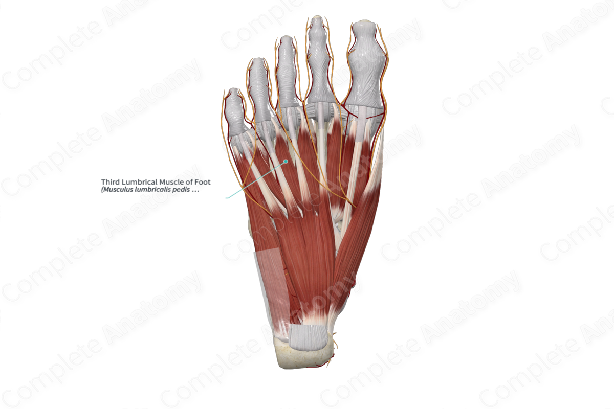
Origin: Tendons of flexor digitorum longus that travel to the third and fourth toes.
Insertion: Medial aspect of extensor expansion of fourth toe.
Action: Simultaneously flexes metatarsophalangeal joint and extends interphalangeal joints of fourth toe.
Innervation: Deep branch of lateral plantar nerve (S2-S3).
Arterial Supply: Lateral plantar artery, deep plantar arch, and plantar metatarsal arteries.


Related parts of the anatomy
Origin
The third lumbrical muscle of foot consists of two heads:
- the medial head, which originates from the lateral aspect of the tendon of flexor digitorum longus that travels to the third toe;
- the lateral head, which originates from the medial aspect of the tendon of flexor digitorum longus that travels to the fourth toe.
Insertion
The fibers of the third lumbrical muscle of foot travel anteriorly to the fourth toe and insert, via a short tendon, onto the medial aspect of the extensor expansion of the fourth toe.
Key Features & Anatomical Relations
The third lumbrical muscle of foot is located in the second layer of muscles that are found in the plantar part of the foot. It is a short, wormlike, bipennate skeletal muscle.
It is located:
- superficial (inferior) to the adductor hallucis muscle and the second plantar interosseous muscle of foot;
- deep (superior) to the plantar aponeurosis;
- medial to the tendon of flexor digitorum longus that travels to the fourth toe;
- lateral to the tendon of flexor digitorum longus that travels to the third toe.
Actions
The third lumbrical muscle of foot simultaneously flexes the fourth metatarsophalangeal joint and extends the interphalangeal joints of the fourth toe (Moore, Dalley and Agur, 2009).
List of Clinical Correlates
- Clawing of the toes
- Charcot-Marie-Tooth disease
References
Moore, K. L., Dalley, A. F. and Agur, A. M. R. (2009) Clinically Oriented Anatomy. Lippincott Williams & Wilkins.
Actions
The third lumbrical muscle of foot simultaneously flexes the fourth metatarsophalangeal joint and extends the interphalangeal joints of the fourth toe (Moore, Dalley and Agur, 2009).
Learn more about this topic from other Elsevier products
Foot Muscle

The extrinsic foot muscles are those whose muscle bellies reside proximal to the foot, but tendons directly insert into the bones and ligaments.
